Black Lives Matter. Supported at Emulate.

Our mission to improve human health is not limited to any race, gender, color, creed, or sexual orientation, and we cannot realize this mission without first acknowledging the inequities in our society and working towards racial equity and justice.

As a company, we believe the ongoing injustice of systemic racism and oppression in our society needs to be addressed. The deaths of George Floyd, Breonna Taylor, Tony McDade, Ahmaud Arbery, and Manuel Ellis—as well as the far too many unnamed Black men and women before them—are a tragedy and a result of centuries-old systemic oppression and racism that have hurt our nation and our Black communities.

As an organization, we stand with many others who honor the Black lives lost due to the devastating effects of racial inequality, racial injustice, and White supremacy. We are also united with common values as part of our company principles called “Being Human”—a set of guidelines created by our team to help us all work in harmony. At its core, “Being Human” is a shared belief that compassion, honesty, and empathy are essential in achieving healthy collaboration toward our mission. But we cannot fully realize this principle without acknowledging unequivocally that Black Lives Matter.

As an organization, we stand with many others who honor the Black lives lost due to the devastating effects of racial inequality, racial injustice, and White supremacy.

We realize that there is work to be done in order to address the rampant inequalities that permeate our society, and we are committing to doing our part.


This includes, but is not limited to:

  1. From June 1, 2020 through December 31, 2020, we are pledging to match employee donations to the below local and national organizations:

  2. We are working to create a more inclusive workplace so all Emulate employees feel safe, comfortable, and valued. This includes:

    • Ensuring our hiring and professional development policies to promote diversity and racial equity across our organization.

    • Implementing anti-racism education and trainings to our entire team with the help of outside diversity and inclusion professionals and anti-racism educators.

    • Ensuring there are anti-bias practices in our hiring process to proactively level the playing field for all candidates.

  3. We are updating our time-off policies to support paid time-off for employees who choose to exercise their constitutional rights to protest or assemble for the sake of any social movement or gathering.  

This is just the beginning. Our work will be ongoing as we continue to learn and grow as an organization, and we are committed to taking action to bring an end to injustice and inequality.


Using a Human-Relevant Model

Shigella are bacteria that cause Shigellosis, a gastrointestinal disease inducing severe stomach cramps, fever, and diarrhea. The onset of symptoms can begin from 1-2 days of contact and results from the rapid invasion of the pathogen within intestinal epithelial cells, leading to the destruction of healthy human colon tissue. In developing countries, Shigellosis remains an important health threat and can lead to epidemics where sanitation is poor[1]. Every year, Shigellosis kills hundreds of thousands of individuals, mostly children younger than the age of five[2].

It is challenging to study this pathogen because Shigella infects only humans, resulting in colon infection. Therefore, small animal models, such as mice, cannot be used to study the disease.  Also, conventional cellular in vitro models, such as human polarized epithelial cells grown on transwells, fail to develop efficient infection. Thus, it has been difficult to study the inception of Shigella infection and understand how this pathogen interacts with the epithelial barrier at the tissue scale. For decades, the scientific community has been missing a reliable, consistent, and clinically relevant Shigella infection in a polarized intestinal epithelial monolayer.

Leveraging Organs-on-Chips technology from Emulate, researchers published a paper in Cell Host & Microbe, highlighting how a human Intestine-Chip can be used to test the infectivity of Shigella. The paper showcases a number of experimental approaches such as real-time imaging, tissue cross-section analysis, and conventional microbiology.

Creating the Intestine-Chip for Shigella Testing

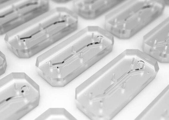
First, the microenvironment of the human intestine was recreated by utilizing Organs-on-Chips technology to create an Intestine-Chip model. The chip itself is made of an elastomeric polymer which consists of a central channel separated horizontally by a flexible porous membrane; two hollow vacuum channels laterally frame the central channel. The application of cyclic vacuum within these lateral channels induces strain and stretching of the porous membrane that recreate peristaltic-like motions (in vivo-relevant mechanical forces). In order to support the culture of human colonic epithelial cells, Caco-2 cells were used, and the separating membrane was coated with extracellular matrix (ECM). To recreate the physiological microenvironment of the intestine, the seeded cells were exposed to continuous flow for 5-6 days to recreate the shear stress on the epithelial channel and cyclic mechanical strain to reproduce the peristaltic motions.

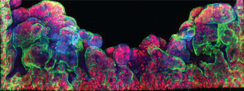
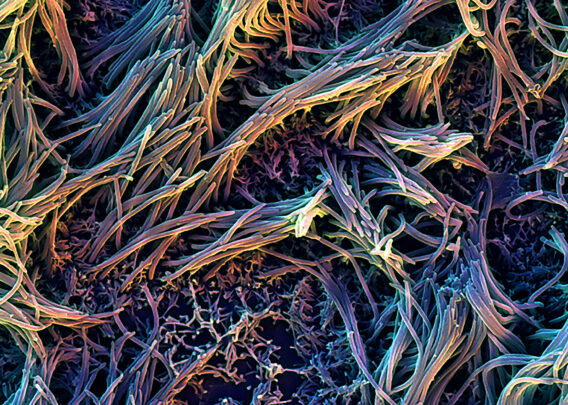
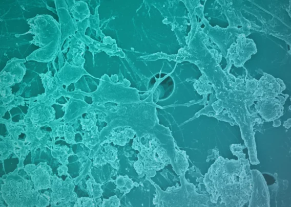
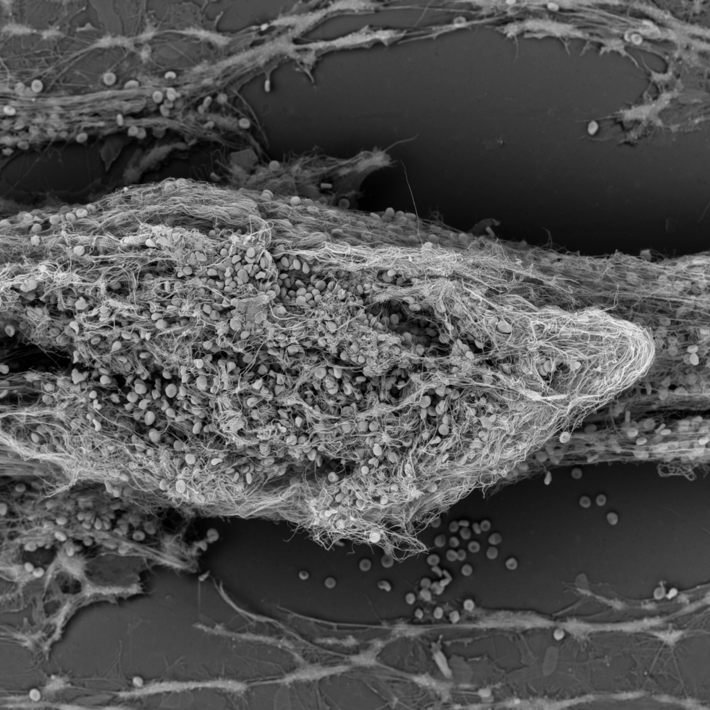
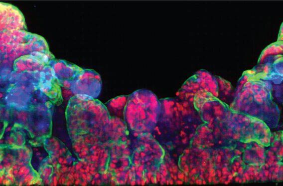
Cells grown under these conditions self-organized into villi-like structures interspaced by crypt-like invaginations. Confirming that the seeded Caco-2 cells were polarized and exhibited known differentiation markers (E-cadherin, villin, and FABP1-KRT20). Scanning electron microscopy (SEM) imaging also showed microvilli in the epithelial channel, a typical structural marker of differentiated and polarized enterocytes.

Next, in order to address whether Shigella flexneri could infect the human colon epithelium in the chip, a wild-type strain expressing GFP (Shigella-WT-GFP) in the intestinal lumen of the Intestine-Chip was introduced.

Human-Relevant Data

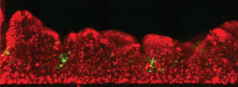
Shigella flexneri distribution within crypt/villi-like formations from the epithelium of the human Intestine-Chip Stitched images showing a cross-section of an Intestine-Chip infected by Shigella-WT-GFP (green) for 1 h. Nuclei were stained with Dapi (red). Scale bar represents 100 mm.

Shigella invasion was observed when only a few hundred bacteria were introduced into the intestinal lumen. The observation that only minimal loads were required for infection is consistent with data from the clinic. Given this correlation between in vivo and chip data, the approach in the chip allowed us to dissect the fundamentals mediating Shigella invasion at the tissue scale. In particular, identification that Shigella directly infect enterocytes from the epithelial channel, shifting the current paradigm about the early stage of invasion. Secondly, the study found that Shigella take advantage of the 3D topology of the tissue to populate specific areas in the colon tissue reminiscent of the human colon crypts. Finally, the design of the Intestine-Chip recreates the mechanical forces on the pathogen invasion and found that peristalsis is critical for specific stages of the infection process.

Most surprising was the observation that Shigella could directly infect enterocytes from the epithelial side (from the intestinal lumen) as expected in vivo with such efficiency and reproducibility.

Collaboration

This work is the result of a collaboration between the Unité de Pathogénie Microbienne Moléculaire and Biomaterials and Microfluidics Core Facility at Institut Pasteur and Emulate. It exemplifies how joining forces from different areas of expertise (engineering, microbiology, and bio-imaging) can reveal new avenues to better understand the mechanism of infection by enteroinvasive bacteria. It also helps advance our understanding of human disease, ultimately enabling the development of new therapeutics.


References:

[1] NIH: https://www.ncbi.nlm.nih.gov/books/NBK8038/

[2] CDC: https://www.cdc.gov/shigella/index.html

Designing the Human Emulation System

The most powerful technologies of the past 15 years have been revolutionary not only because of what they enable people to do, but also because they are easy-to-use.

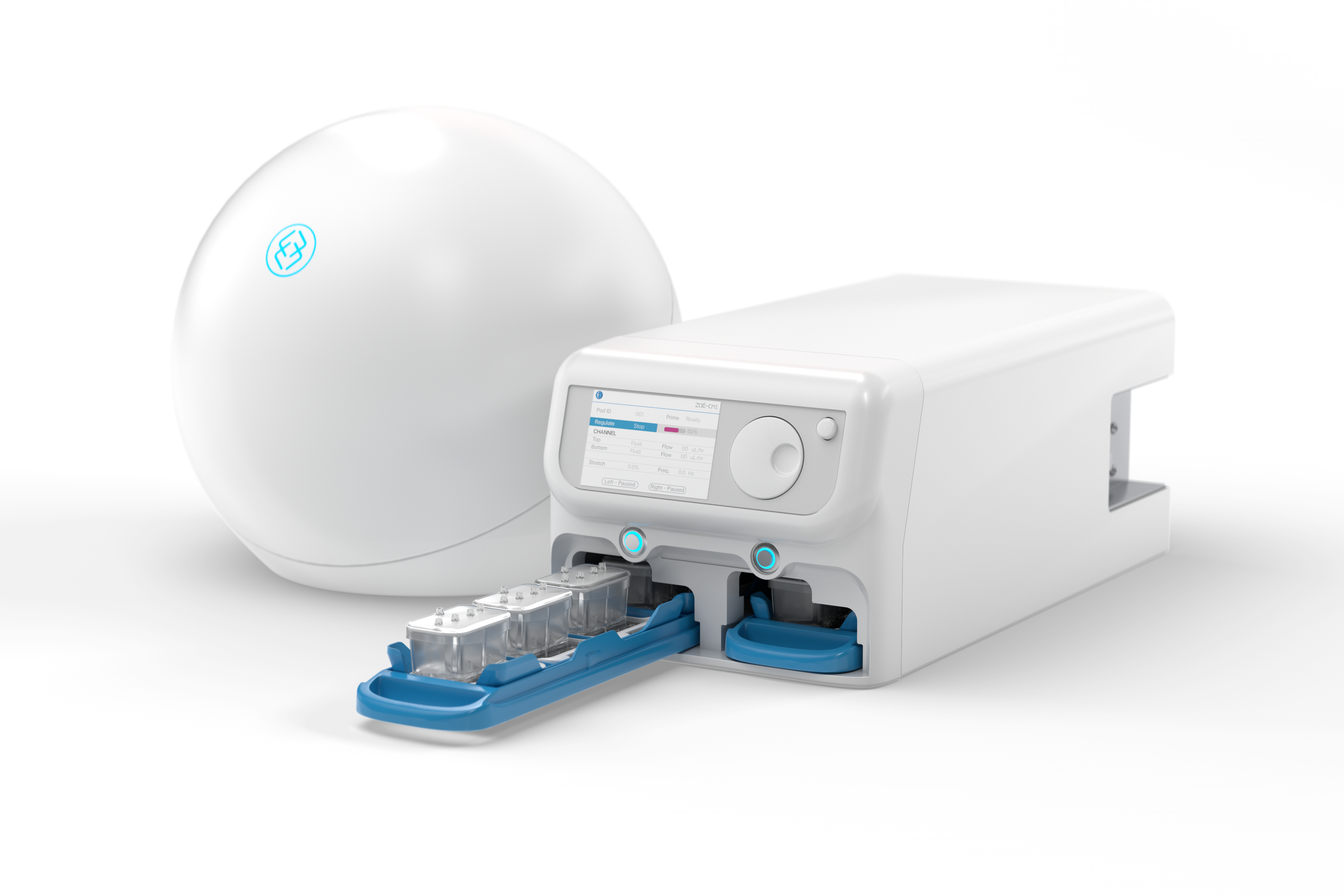
We designed our Human Emulation System to embody this philosophy. It takes Organ-on-a-Chip technology—which has traditionally required specialized engineering knowledge—and integrates it with hardware and software apps. This Organ-Chip technology platform can be utilized in contemporary laboratory workflows and provides advanced automation so that researchers can run experiments repeatedly and robustly in a variety of environments.

The development of the Human Emulation System started with the creation of our first Organ-Chips at the Wyss Institute of Biologically Inspired Engineering. At the time, we knew that an Organ-Chip solution held enormous potential to provide a better understanding of human biology and disease. But we also knew that we had to build an Organ-Chip technology platform that users with different training could use in their labs.

Chip-S1™ Stretchable Chip

Organ-Chips are living, micro-engineered environments that recreate the natural physiology and mechanical forces that cells experience within the human body. They recreate human biology more accurately than animal models or other cell culture methods. Our Chip-S1™ Stretchable Chip—our first commercially available chip design—can be configured to emulate different organs, including lung, liver, intestine, kidney, and brain.

The microenvironment created within each Chip-S1 includes epithelial cells in the top channel and endothelial cells in the bottom channel. The top and bottom channels are separated by a porous, flexible membrane that allows for cell-to-cell communication that is similar to those that are found in the body. These two channels are fluidically independent and users can determine the rate of flow through the channels.

Pod-1™ Portable Module

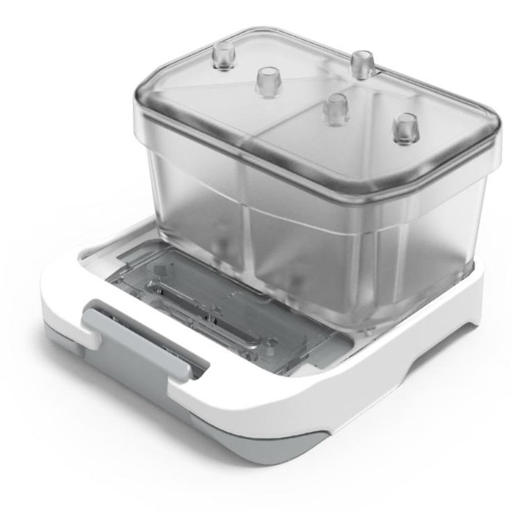
Our Pod houses the chip, supplies media, and enables compatibility with microscopes and other standard laboratory analysis equipment. The design of the Pod allows Organ-Chips to be easily transported around the lab and placed on microscopes for imaging. The Pod’s reservoirs allow users to introduce nutrient media, precisely control dosing to test drugs or other compounds, and sample chip effluent.

Zoë-CM1™ Culture Module

Our Zoë Culture Module is designed to sustain the life of cells within our Organ-Chips. It provides the dynamic flow of media and the mechanical forces that help recreate the microenvironment cells experience in vivo. The instrument automates the precise conditions needed for simultaneous cell culture of up to 12 Organ-Chips.

The culture module enables independent control the flow rate of media through the top and bottom channels of the chips, as well as stretch parameters — including frequency and amplitude. Additionally, Zoë has automated algorithms to prime the fluidic channels of Pods with media, and programming to maintain the culture microenvironment for optimal cell performance.

Orb-HM1™ Hub Module

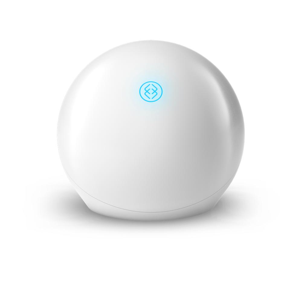
The Orb Hub Module provides a simple solution for installing and operating the Human Emulation System within the lab environment. It delivers a mix of 5% CO2, vacuum stretch, and power from standard lab connections. It generates the 5% CO2 supply gas to Zoë by combining air with an external 100% carbon dioxide supply or from a portable CO2 canister.

Software Apps

Our team is developing a suite of apps that can be used to plan and execute experiments, analyze data, and foster collaboration between and among teams. This shared ecosystem of apps is also designed to help teams write, edit, and share protocols and standardize the data they create. What’s more, these apps help users visualize and interpret datasets to improve an understanding of human biology and disease.

RECREATING DRUG SIDE-EFFECTS USING ORGANS-ON-CHIPS

Maintenance of the fluidity of blood within the circulatory system is an important physiological process that is tightly regulated by the endothelial cells that line the blood vessels of the human body. The term thrombosis usually refers to pathological blood clotting that can manifest clinically in relation to dysfunction of the circulatory system. As more therapeutics have been developed, it has also become apparent that some drugs can contribute to the development of thrombosis, resulting in serious adverse events.

It is difficult, however, to predict drug-induced thrombosis using current pre-clinical cell culture and animal models, due to the dynamic nature of the flow of blood in the body and the complex molecular mechanisms that regulate drug interaction with different blood components of the human circulatory system.

Human-Relevant Models

In a paper published in Clinical Pharmacology and Therapeutics, a Blood Vessel-Chip was developed to test a compound, a monoclonal antibody called Hu5c8, that had been shown to cause unwanted thrombosis in humans. Hu5c8 displayed promise as a treatment for lupus, but was ultimately terminated during clinical trials due to unexpected thrombotic and cardiovascular events it caused in patients. Unfortunately, these life-threatening side effects were not discovered during preclinical testing due to a lack of human-relevant models that could predict them. Using the Blood Vessel-Chip, researchers detected for the first time the pro-thrombotic effects of Hu5c8 using one clinically relevant dose of this compound in combination with human blood samples, and the data generated were consistent with previous clinical findings.

What’s perhaps even more interesting is that although previous studies have shown that thrombosis can be recreated in vitro, these results were limited to imaging data, since the formation of the clot made it impossible to obtain soluble samples. But in recent work, a team of researchers created a new chip design that enabled the ability to sample and analyze biomarkers from a clotted chip, allowing insight into mechanism of action of how Hu5c8 led to thrombosis.

Efficacy Testing of Anti-Platelet Drugs

With this chip design, researchers could induce thrombosis by stimulating endothelial inflammation, or by adding blood-borne, pro-coagulant factors. This approach created the ability to study the efficacy of anti-platelet drugs and is highlighted in the paper published in collaboration with Janssen Research & Development, the Wyss Institute at Harvard, and the University of Twente. This research shows how Organs-on-Chips technology enables the use of human-relevant models of drug-induced thrombosis, helps us dissect the molecular mechanisms that mediate this process, and it could also be used to study the efficacy of new anti-thrombotic therapeutics.

A New Initiative Using Organs-on-Chips Technology

Researchers know that people respond differently to foods, drugs, and chemicals. For example, a medication may be effective in one individual, while it makes another even more ill. Until now, we’ve had no way to test how a diet or a treatment plan would affect a person before administering it to them.

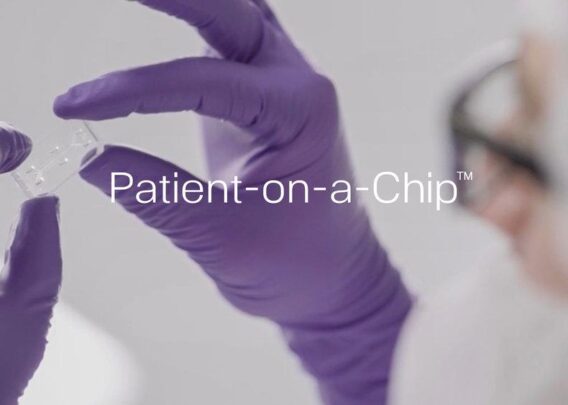
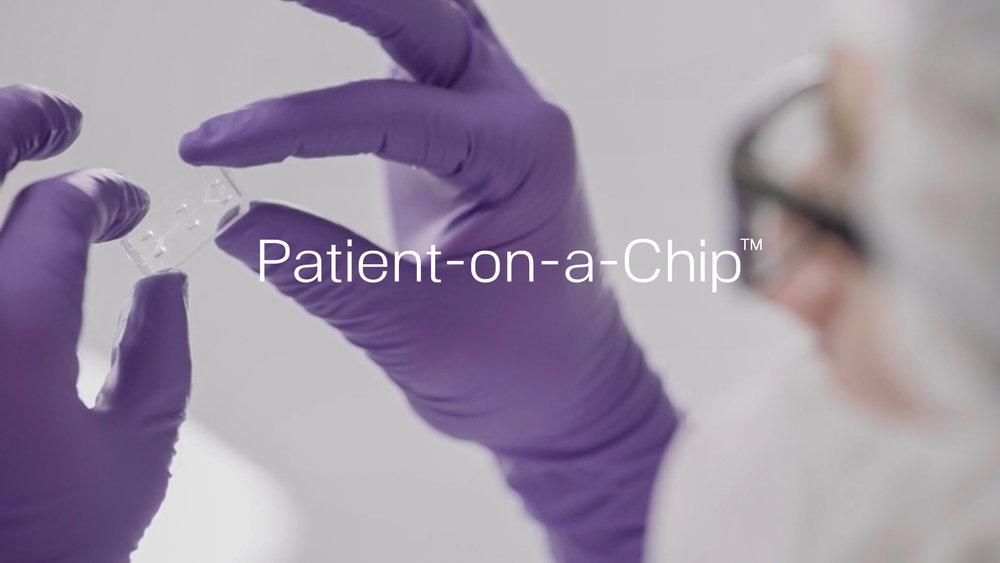
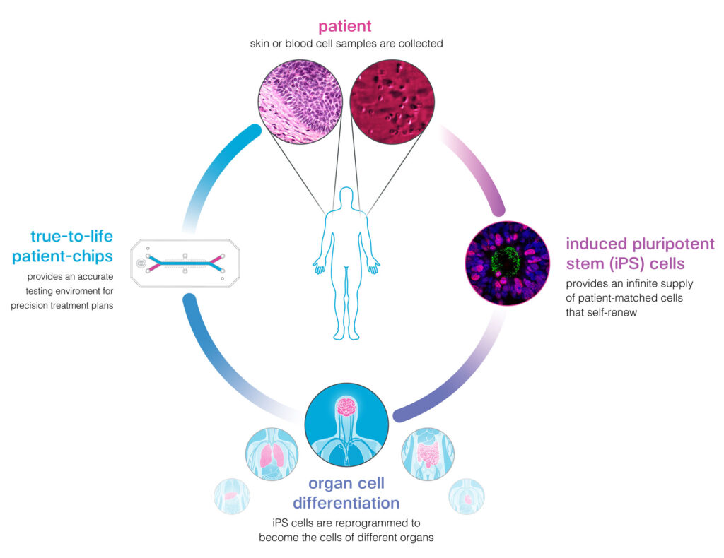
In an effort to provide clinicians with better information about their patients, Emulate has an ongoing program with Cedars-Sinai Medical Center that combines their expertise working with stem cells together with Organs-on-Chips technology. This Patient-on-a-Chip program holds the potential to advance the field of precision medicine and provide clinicians with a new system that more accurately predicts an individual’s response to foods, medicines, and chemicals.

Combining Stem Cells and Organs-on-Chips

The collaboration leverages innovative stem cell science from Cedars-Sinai and the Human Emulation System, which uses Organs-on-Chips technology to recreate true-to-life biology outside the body. The Human Emulation System creates an environment where cells exhibit an unprecedented level of biological function and gives researchers the ability to control complex human biology and disease mechanisms that are not possible with other techniques.

Cedars-Sinai scientists can harvest cells from the blood or skin of participating individuals. These cells can then be reprogrammed into induced pluripotent stem cells, which can be made into any organ cell, each bearing the unique genetic fingerprint and characteristics of the individual. This process is particularly useful to recreate cells from organs like the brain, which can’t be sampled from a living individual.

By placing a patient’s cells in Organ-Chips and exposing those cells to a particular drug or series of drugs, clinicians could gain better information about how that individual would respond to treatment, avoiding the risk of administering a drug that may cause harm.

Researchers at Cedars-Sinai can turn cells from a sample of blood or skin into induced pluripotent stem cells, and then reprogram those cells into different organs cells found in the body. These organ cells can then be put on our chips.

The Promise of Precision Medicine

The promise of precision medicine is that when a patient is sick, a clinician will be able to develop a tailored treatment plan that takes into account that patient’s unique biology. Organs-on-Chip technology will provide better information about how a patient will respond to a disease and to a subsequent treatment that will help doctors make more informed decisions.

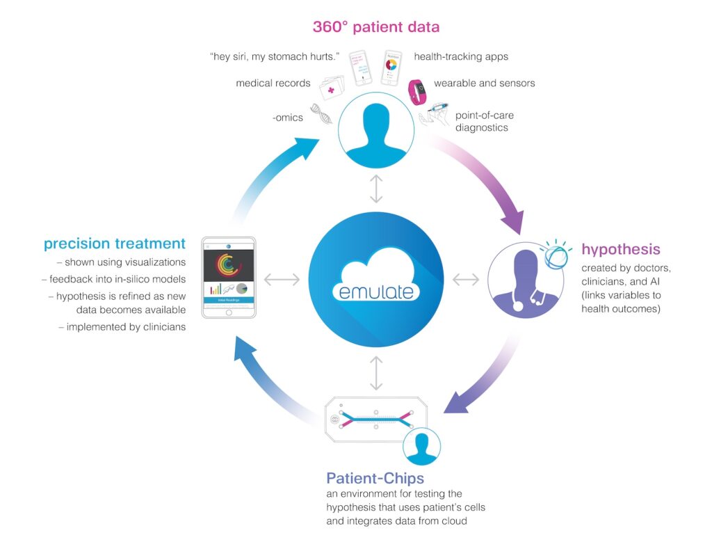
As science continues to advance, precision medicine will include preventative measures as well. Imagine a recursive circle of precision medicine that integrates the expertise of doctors and clinicians, data generated by wearables and other devices, the analytic and predictive power of artificial intelligence, together with Organs-on-Chip technology.

In the recursive circle of precision medicine, the expertise and experiential knowledge of doctors and clinicians will be supplemented with data produced by our living products and the analytic power of AI.

Transforming Care

This project holds the potential to change the way diseases are treated. It will also change the way each of us understands and manages our own health by providing deeper insights into our own biology.

“Cedars-Sinai’s world-class stem cell expertise and discovery, combined with the pioneering Human Emulation System, is poised to reshape the future of medical care,” said Shlomo Melmed, MB, ChB, executive vice president, Academic Affairs, and dean of the medical faculty at Cedars-Sinai.

Initial scientific findings, have been published in Cellular and Molecular Gastroenterology and Hepatology, a journal of the American Gastroenterology Association.

Organ-Chips in Space

Increasing Our Understanding of the Effects of Space on the Human Body

The human body was not designed for space. Low gravity causes muscles to waste away and bones to lose their density. Without the protection of the Earth’s magnetic field, cosmic rays and solar radiation bombard tissue and cells, inflicting damage that could cause disease.

Living out there is psychologically taxing as well. Without the Earthly cycle of night and day, astronauts struggle to maintain their natural sleep schedule, which can lead to changes in mood and mental performance — never mind simply being so far from home, trapped in a living area the size of minivan, with only a few other folks to talk to for months on end.

Still want to volunteer for that nine-month trip to Mars?

Space agencies have designed ways to replicate the condition of low gravity on Earth, but this is only an approximation of the effects of space on the human body and how it will respond. The only way to figure out how an astronaut will react is to put them on a rocket and shoot them into orbit.

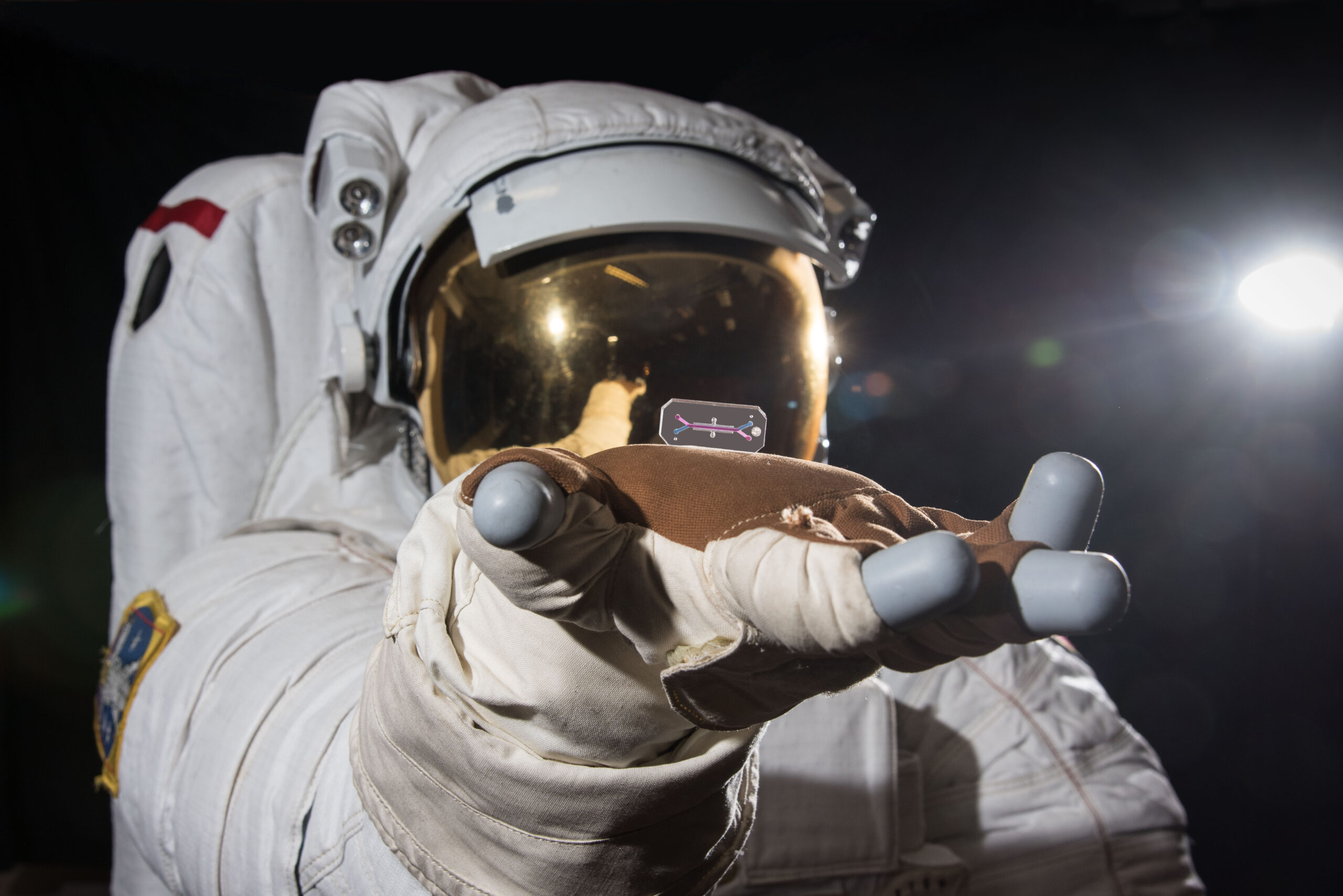
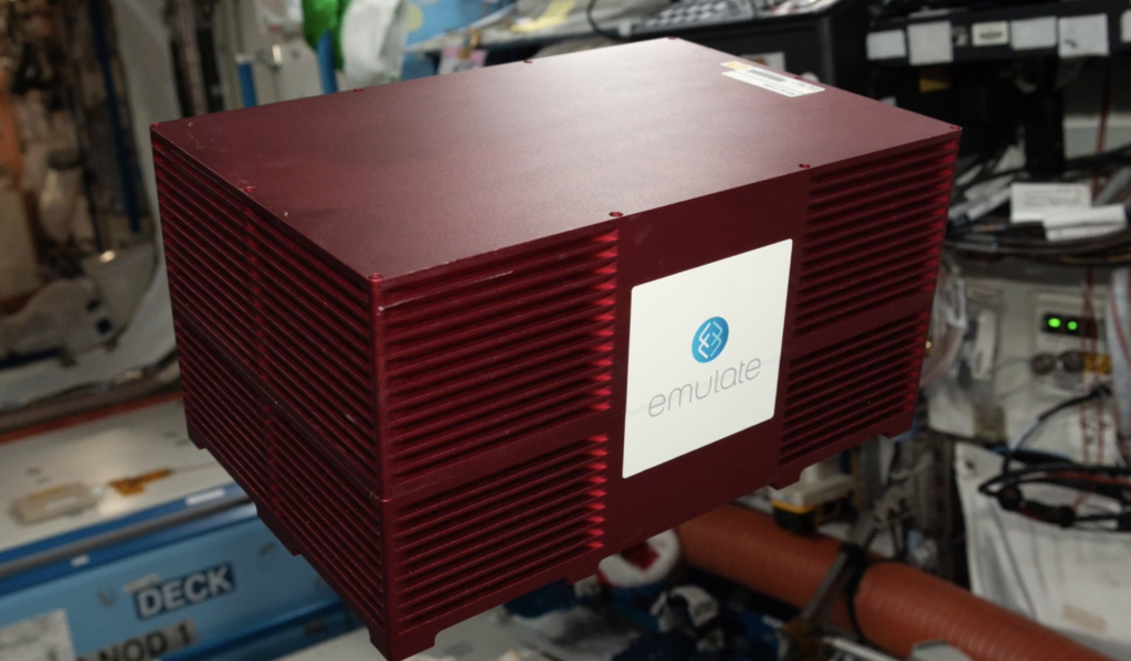
As an alternative, we’ve teamed with NASA and IRPI, a scientific consulting firm, to send our Human Emulation System to the International Space Station (ISS). The goal is to test the cells of astronauts using Organ-Chips in space, providing data about how their body reacts.

Personalized (Space) Medicine

The future of our Organs-on-Chips technology lies in personalized medicine, or the ability to tailor preventative and therapeutic treatments to an individual’s genetics and life experiences. If we could take an individual’s cells and put them on Organ-Chips, we could get an accurate sense of how medicines, foods, and chemicals would affect that person as an individual.

We could then take this process one step further by collaborating with NASA to develop Organ-Chips from the cells of astronauts. Imagine a scenario where an astronaut is in training for a long mission in space. We could take a blood sample from this astronaut, extract stem cells found in their blood, and then engineer those stem cells into different organ cells. This would allow us to create Organ-Chips with DNA that matched the individual astronaut. We could then send those Organ-Chips to space and run experiments on our Human Emulation System aboard the ISS to see how those cells responded in low Earth orbit.

“Eventually, we could send an astronaut’s cells to the ISS before we send him or her up there, getting a sense of how their cells will react to low earth orbit and all the environmental stressors that come along with that,” said Chris Hinojosa, VP, Platform Development at Emulate.

Pushing the Limits of Organs-on-Chips

Nasa Launch

We are democratizing our Human Emulation System by making it easy to use in different kinds of labs throughout the world. But a trip to space could be seen as the ultimate test of our logistics and our platform.

A huge amount of planning goes into launching a rocket, whether it is manned or unmanned. If we can get our Organ-Chips and our lab-ready Human Emulation System into orbit so that they can be used for significant experiments, we’re confident that we can get them to any lab on Earth.

“Pushing the limits of our technology will encourage us to innovate even more, and this kind of work will have a huge impact on our technology,” Hinojosa said.

Deeper into the Unknown

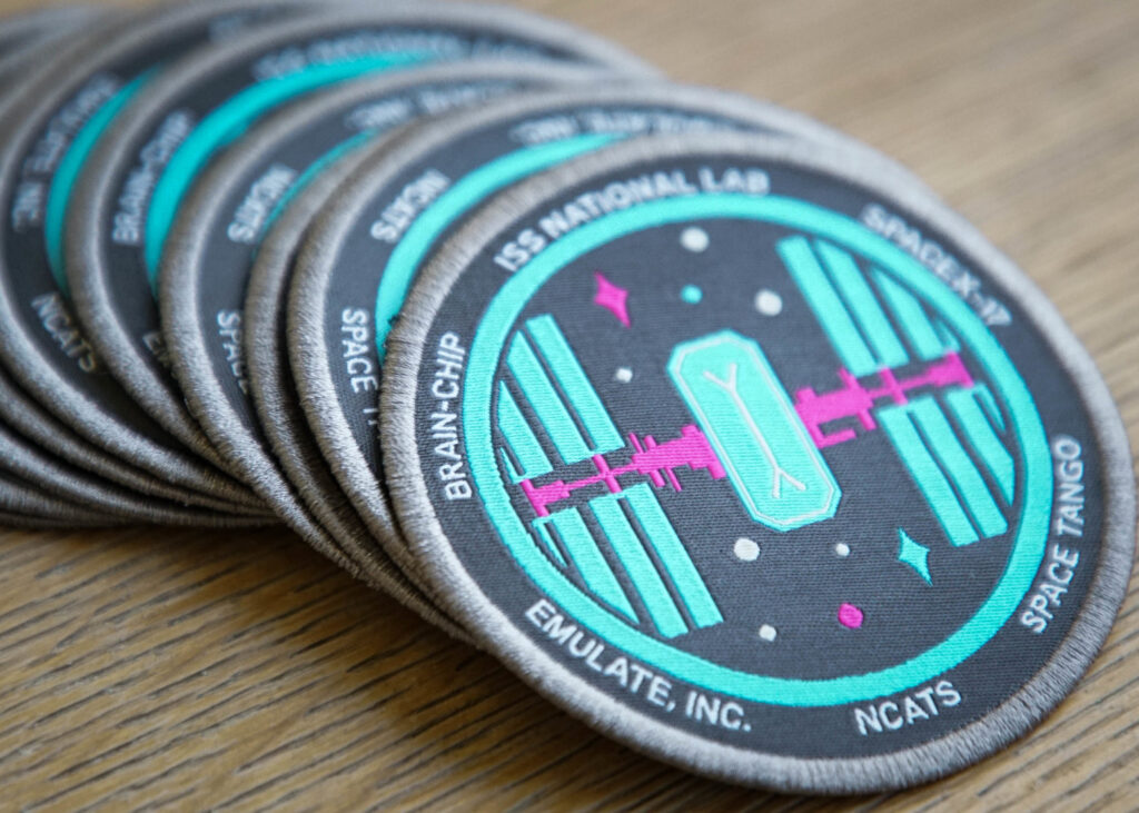
ISS Patch

The ISS circles the Earth at an altitude of approximately 200 miles. As far away as it may seem, the station is still sheltered by a portion of the Earth’s magnetic field, which protects the ISS and the astronauts inside from harmful radiation.

Rarely have humans traveled outside the Earth’s magnetic field. Indeed, the last manned flight outside Earth’s orbit was the final Apollo mission in 1972. And because humans have made so few voyages outside the magnetic field, we simply don’t know how the human body will respond to an extended period beyond its protection.

In the coming decades, we are likely to go deeper into the cosmos than we ever have before. Though still in the planning stage, NASA and private space agencies are working on manned missions to Mars.

The scientific community has a sense of how interplanetary travel will affect human biology. But perhaps our Human Emulation System could be the first human cells to be sent beyond the moon — and provide scientists with information about how to protect astronauts before the first manned Mars mission launches. More about this project can be found on NASA’s website.