CEO, Jim Corbett, shares how new funding will enable Emulate to expand research and development efforts to new heights

In March 2020, I was fortunate to join Emulate, intrigued by one of those rare opportunities to bring truly game-changing technology to healthcare. 

Five days later, the world shut down.  

Every day since has been motivated by a vital mission to leverage human biology and technology to ignite a new era in human health, driven by a team of people with an unwavering commitment to bring a human-centric approach to biological innovation.

As we were coming to terms with our new normal, our team’s focus and action were nothing short of miraculous. Undaunted, and united under a single vision, the team persevered. We found new ways to move forward with a collective strength to overcome challenges. We embraced new processes to shatter unexpected roadblocks. And we responded to the needs of our customers to ensure their science could keep moving forward.  

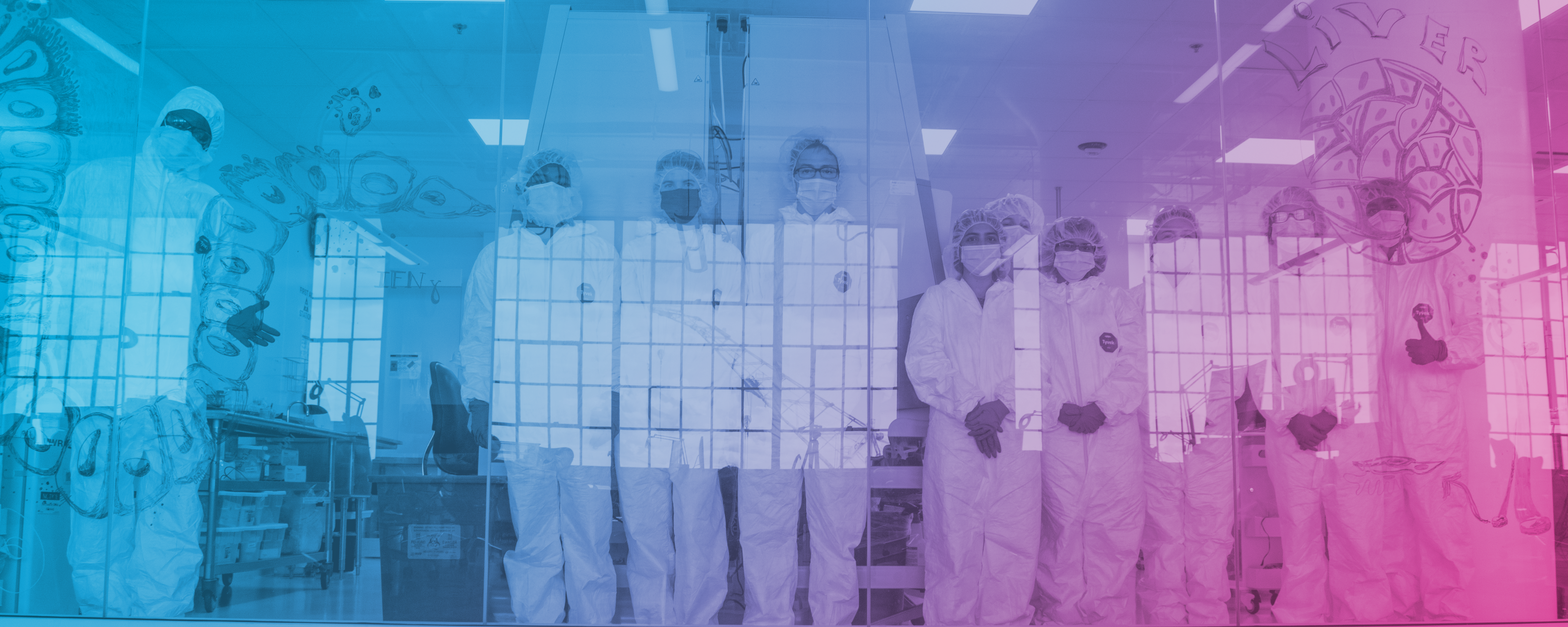
ONE Curious, Brave and Human Team 

We grew our team, adding deep commercial and functional expertise to implement structure and processes in order to rapidly scale. The scientific team has grown to include researchers focused on our application roadmap areas such as immunology and microbiome. We expanded our leadership team with Donald Ingber, MD, PhD, a scientific founder of Emulate and Chairman of its Scientific Advisory Board – and arguably one of the great pioneers in the field of biologically inspired engineering – joining our Board of Directors. Also, Scott Kantor accepted the role of Chief Financial Officer and Larry Weiss came on as Chief Legal Officer.  

Breakthrough Innovations that Challenge the Status Quo 

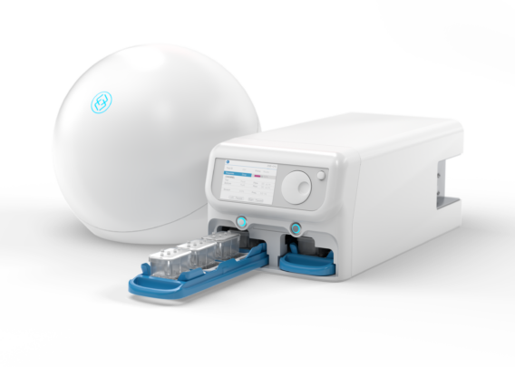
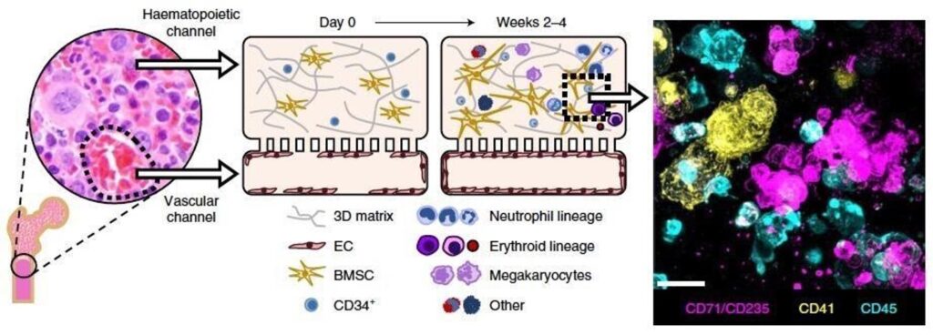
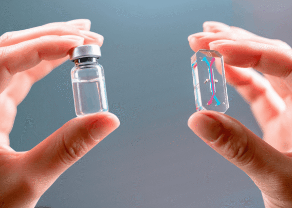
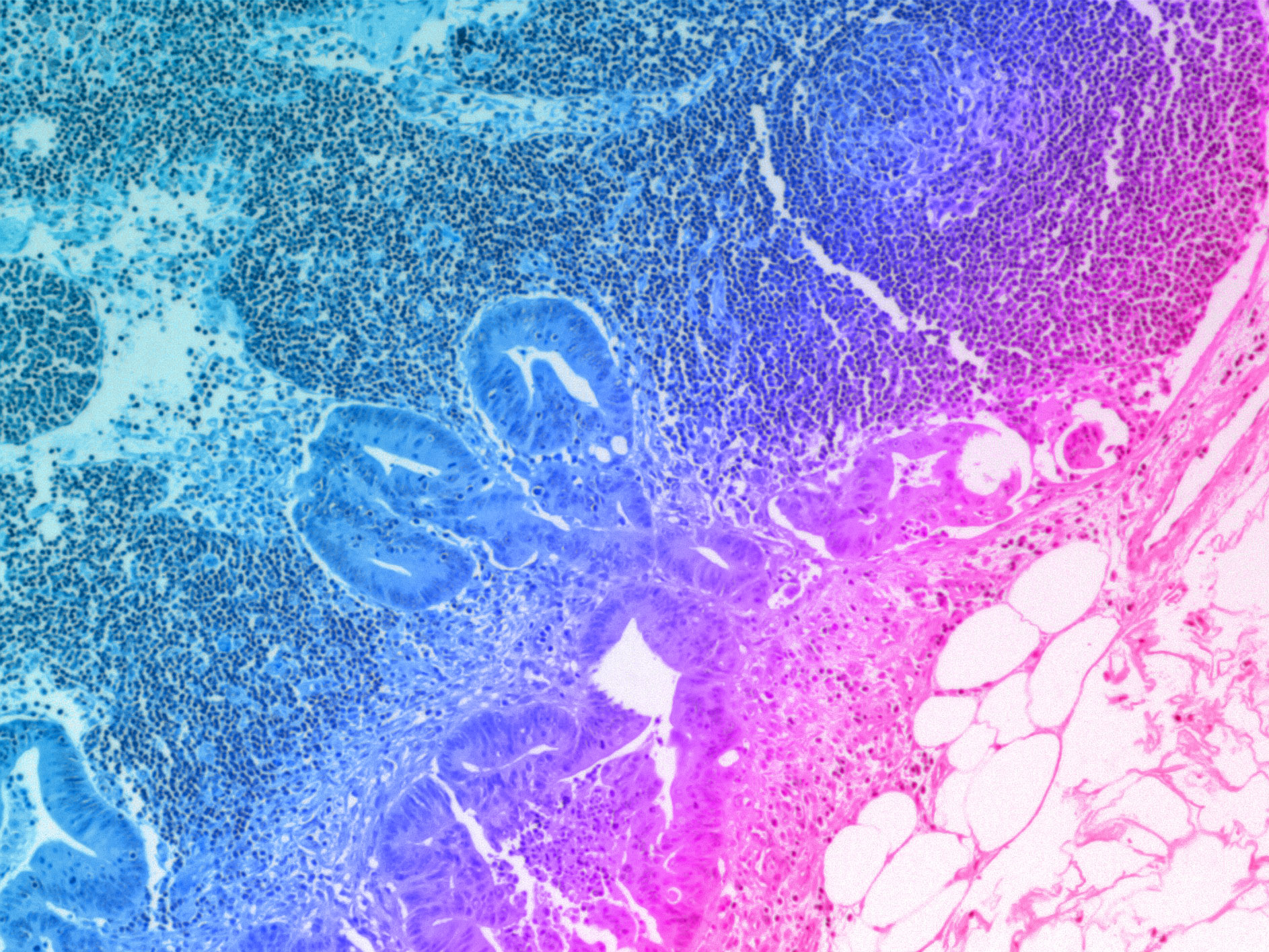
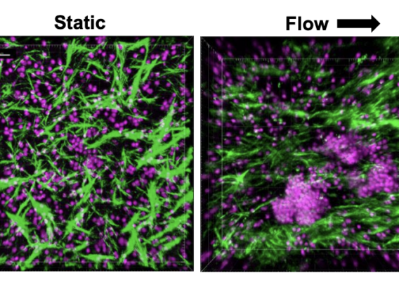
Over the past 18 months, we announced our roadmap for expanding Organ-Chip applications with a focus on developing targeted and advanced biologics. Additionally, we released a series of breakthrough applications and organ-models, such as the Brain-Chip and the Colon Intestine-Chip, deepening our portfolio of the human-relevant Organ-Chips that helped solidify our commitment to scientific excellence. 

We have bolstered our leadership team, accelerated product development goals, and seen healthy growth in demand for our products. Several leading indicators validate our belief that organ-on-a-chip technology will dramatically transform the entire drug discovery and development pipeline and ultimately eliminate unnecessary animal testing.

Jim Corbett, Emulate CEO
Policy-Changing Influence and Advancements 

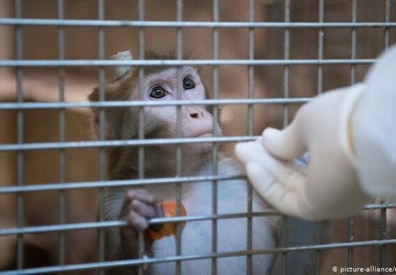
Aside from our product and commercial developments, we have taken a stronger stance against animal testing, as there is scientific proof that it has significant  limitations in effectiveness – particularly for today’s novel drugs and vaccines. In response to a New York Times article, we championed legislation reducing the use of primates for drug testing. More recently, the FDA Modernization Act and the Humane Research Act have been introduced to Congress to rally support for alternatives to animal testing, citing scientific inadequacies and ethical concerns associated with it. Our team also joined the North American 3Rs collaborative and believe that moving in the right direction means encouraging the use of more accurate and humane technologies. We truly believe that organ-chips will become an integral part of drug development and has the potential to ultimately eliminate animal testing.

Onward Movement 

This level of achievement would be astonishing in normal times and no doubt helped propel our fundraising efforts. Today, we announced the close of an $82 million dollar round of financing, which will enable Emulate to expand research and development efforts to new heights. We will scale-up R&D activities to facilitate the creation of new human-relevant organ-on-a-chip models in immunology, neuroinflammation, tumor modeling, and more. To help meet growing global demand, we will extend operations in the Asia Pacific region with new distributor relationships. 

I am excited to see what this team will accomplish as we set out into a changed world, continuing to scale and meet global demand as well as allow researchers to predict human response better than conventional cell culture and animal-based experimental testing. This biological revolution is just getting started. Let’s go! 

A summary of the paper published in Nature Biomedical Engineering: On-chip recapitulation of clinical bone marrow toxicities and patient-specific pathophysiology.

Human bone marrow is the body’s major site of hematopoiesis, the proliferation and differentiation of all blood cellular components from hematopoietic stem cells. These actively dividing and maturing cells are highly sensitive to toxic stimuli and, thus, bone marrow is one of the major targets of unwanted toxicity caused by drugs or therapeutic agents. Predicting drug-related hematotoxicity with clinically relevant pharmacokinetics would be extremely valuable for designing safety assessments and human trials. 

Studying bone marrow tissue is particularly difficult due to its inaccessibility and the limited number of physiologically relevant in vitro models that can support growth and maintenance of bone marrow cultures. Current hematopoietic toxicity studies are performed using methylcellulose colony cultures in which cells are suspended and can be exposed to higher-than-usual concentrations of drugs for extended periods of time. To address the unmet need for a predictive model to study hematopoietic dysfunction and pathophysiology of bone marrow, the authors created a Bone Marrow-Chip (BM-Chip) to recapitulate relevant hematopoietic cell types in a three-dimensional (3D), mechanically active next-generation in vitro model. This model possesses relevant levels of complexity to closely mimic the in vivo environment to examine the effects of known myelotoxic stressors and to investigate a genetic condition resulting in bone marrow failure.

  • Research Area: Drug development, Toxicity testing, Rare Disease
  • Organisms: Human
  • Sample Types: Human bone marrow-derived mesenchymal stromal cells, hematopoietic stem/progenitor (CD34+) cells, umbilical cord vascular endothelial cells
  • Research Question: Can a Bone Marrow-Chip recapitulate the features of human hematopoiesis and dysfunction resulting from genetic mutations and exposure to drugs and radiation?


Experimental Overview

Characterizing the Bone Marrow-Chip

Researchers created the BM-Chip using human bone marrow-derived stromal cells (BMSC) and CD34+ cells co-cultured in a fibrin gel in the upper channel of the chip. The lower channel of the chip contained human umbilical vein endothelial cells (HUVEC) with unidirectional continuous flow of culture medium containing cytokines that support cell growth (stem cell factor, Flt3 ligand and thrombopoietin) as well as factors that encourage multilineage differentiation (granulocyte colony stimulating factor and erythropoietin). Compared to the same cells grown in static models in suspension and in 3D gel, the BM-Chip increased the numbers of proliferating myeloerythroid and neutrophil progenitors, demonstrating increased cell survival and robust hematopoiesis in vitro over prolonged culture time (28 days).  

Schematic of bone and insert micrograph showing normal human bone marrow histology. Left middle: schematic of the cross-sectional view of the human BM-Chip on day 0 after seeding, showing singly dispersed CD34+ progenitors and BMSCs in a gel in the top channel and an incomplete vascular lining (seeded on either day 0 or day 8) in the bottom channel. Right middle: within 2 weeks of culture initiation, endothelial cells grow to cover all four sides of the lower channel and create a vascular lumen, while CD34+ cells undergo expansion and multilineage differentiation. Right: immunofluorescence image of a vertical cross-section through the gel in the upper channel of the BM-Chip taken at day 14 (magenta: erythroid lineage; yellow: megakaryocyte lineage; blue: neutrophil and other hematopoietic lineages). 

Modeling Drug-Induced Hematotoxicity in the Bone Marrow-Chip

To determine if the BM-Chip could be used to model human bone marrow pathophysiology, the researchers exposed the chips to 5-fluorouracil (5-FU) at dosage-appropriate plasma concentrations to observe toxic effects. BM-Chips from six different CD34+ donors infused with 5-FU through the vascular (bottom) channel consistently showed hematotoxicity at clinically relevant concentrations, whereas suspension and static gel co-cultures did not.

In the next set of experiments, the researchers challenged the BM-Chip with AZD2811, a small-molecule inhibitor of Aurora B kinase that is critical for proper cell division and is currently in phase II clinical development as a cancer therapeutic drug. In earlier phases of clinical trials, AZD2811 has been shown to selectively target dividing neutrophil and erythroid cells with varying effects in a time and dose-dependent manner. The authors used the BM-Chip to explore these effects on both mature and progenitor cell types and provide insight into the drug’s mechanisms of action. 2- and 48-hour AZD2811 infusions resulted in dose-dependent toxicities of the erythroid lineage, with much greater effect following the 2-hour infusion regimen than the 48-hour regimen. In the neutrophil lineage, dose-dependent toxicity in the BM-Chip was greater with the 48-hour infusion. Importantly, both regimens revealed maturation-dependent sensitivity to the treatment via selective decrease in immature cells of both lineages compared to mature cells.

Suspension cultures of CD34+ cells failed to replicate the dose-dependent erythroid toxicity that was successfully modeled by the human BM-Chip, suggesting that it can more accurately predict hematological effects at clinically relevant drug exposures than static culture models.

Radiation and Bone Marrow Recovery

Building on the observation that immature, proliferating cell types were selectively affected by AZD2811 treatment, the authors tested maturation-dependent toxicity caused by ionizing radiation. Radiation treatments are widely used as a cancer therapy, and they commonly cause bone marrow depletion – a reduced production of blood cells from all lineages. The authors noted that the BM-Chips showed decreased numbers in all cell types studied that correlated with increasing doses of radiation. These experiments show that the BM-Chip is a useful model of human-specific radiosensitivity, inviting more detailed analyses of radiation-associated cell death.

The researchers went on to demonstrate that the BM-Chip can also be used as an in vitro model to examine bone marrow recovery and response to potential therapeutics using human-relevant pharmacokinetic profiles during preclinical drug evaluation, development, and regulatory assessment, which cannot be done using animal models.

Modeling Rare Conditions: Shwachman-Diamond Syndrome

Shwachman–Diamond syndrome (SDS) is a genetic condition that causes bone marrow depletion and failure, resulting in neutropenia. The extent of hematological dysfunction may involve other cell lineages as well, leading to myeloid malignancies in some SDS patients. Understanding SDS has been limited by a lack of animal models that faithfully recapitulate the condition. In this study, the authors cultured CD34+ cells from two patients with SDS alongside normal BMSCs and endothelial cells to understand the clinical manifestations of the syndrome using this disease model.

The researchers noted that the cells in the SDS BM-Chips exhibited defective hematopoiesis, with fewer numbers of neutrophils and erythroid cells, as well as impaired maturation of neutrophils. The blunted maturation of neutrophils, denoted by their pattern of CD16/CD13 expression, matched retrospective patterns found in bone marrow aspirates of other SDS patients, suggesting that the BM-Chip can replicate the phenotype and some features of the disease. Further studies on the mechanisms of bone marrow failure, anemia, and neutropenia should be possible using the SDS BM-Chip.

Conclusions

This study provides evidence that the human BM-Chip is an effective preclinical model of human BM pathophysiology, enabling analysis of human responses to clinically relevant drug pharmacokinetics profiles and radiation dose exposures. The data show that the BM-Chip supports erythroid differentiation as well as myeloid development and mobilization over four weeks of culture while improving the maintenance of CD34+ progenitors compared to traditional culture methods. Additionally, the BM-Chips were able to better recapitulate the toxicity responses of human bone marrow to clinically relevant dose exposures of AZD2811 and 5-FU and showed an improved ability to recover after injury. 

BM-Chips derived from patients with Shwachman-Diamond syndrome showed dysfunctions that parallel key hematological abnormalities observed in vivo and led to the discovery of a neutrophil maturation abnormality that exists in a significant subset of SDS patients. Taken together, next-generation in vitro modeling of human bone marrow using Emulate’s microfluidic organ-chips has great potential to facilitate drug development, disease modeling, and translational studies for a range of hematopoietic disorders and toxicities – all while offering a human-specific alternative to animal testing for regulatory assessment. 

Testing new vaccines against seasonally emerging influenza strains is a necessary process, due to frequent changes in the virus’s immunogenic surface proteins. Vaccines and other drugs, such as immunotherapies, are evaluated for efficacy and safety in vitro using cultured human immune cells and in animal models. However, these assessments often fail to fully predict human responses, because they lack the biologically relevant microenvironment in which to develop an adaptive immune response. 

Modeling a true human immune response is a complex undertaking. Primary lymphoid organs, like the thymus and bone marrow, allow immune stem cells to proliferate, differentiate, and mature. Secondary lymphoid organs like the lymph nodes, spleen, and tonsils maintain mature but naïve cells ready to be activated by contact with antigen presenting cells, leading to clonal expansion and affinity maturation for the adaptive immune response. Lymphoid follicles (LFs) that are present within lymph nodes or form ectopically within other secondary lymphoid organs contain germinal centers that support the development of plasma cells that secrete high affinity antibodies. 

A human model of the LF would provide a better understanding as to why these organ structures are critical for developing adaptive immune responses and would be extremely useful for preclinical assessment of vaccine efficacy. This study represents the first-of-its-kind human LF-Chip containing B, T, plasma and antigen-presenting cells to test functional immunization responses to vaccines and adjuvants in vitro. 

  • Research Area: Vaccine and adjuvant testing
  • Organisms: Human
  • Sample Types: Peripheral blood-derived immune cells
  • Research Question: Can the LF-Chip be a useful human-relevant preclinical tool for assessing the efficacy and safety of influenza vaccines and adjuvants?


Experimental Overview

Characterizing the LF-Chip

Researchers created the LF Organ-Chip by using peripheral blood mononuclear cells (PBMC) isolated from donor blood and culturing the cells at high density within the extracellular matrix (ECM) in the lower chamber of the chip, with culture medium flow supplied in the upper chamber. The researchers observed self-aggregation of lymphocytes within the ECM resembling which resembled germinal centers. They did not see autoactivation of B cells (determined by levels of IgG or IgM in the Organ-Chip cultures), overcoming a key challenge seen in static 2D cultures. 

Within the LF-like structures formed in the chip, the researchers detected molecules that indicate immune reaction, including the chemokine CXCL13, which recruits T cells, and activation-induced cytidine deaminase (AID) expression, which is required for the critical antibody class switching step. Class switching and plasma cell formation were observed, and, when exposed to an antigen that mimics the presence of dead bacteria, a robust IgG response was generated, suggesting that the model faithfully recapitulated a functional adaptive immune response.

Vaccination Testing on the LF-Chip 

Protective immunity induced by vaccination requires plasma cell production of antigen-specific antibodies. To see if the model could simulate an immune response to vaccination with viral antigens with or without an immune stimulating adjuvant, the authors added patient monocyte-derived dendritic cells in a 3D ECM gel within the Organ-Chip and tested it with influenza virions and adjuvant. The germinal centers showed plasma cell formation, expression of AID by resident B cells, and production of cytokines, and robust production of antibodies to influenza virus hemagglutinin – similar to vaccinated humans. Notably, these results were not replicated within a 2D static cell culture model. A commercial influenza vaccine was used to induce a clinically relevant immune response in the chip, showing important features like cytokine response and donor variability. 

Towards a Better Preclinical Assessment of Vaccines and Adjuvants

This is the first reported in vitro model that supports formation of human LFs with functional germinal centers similar to those found in secondary lymphoid organs in vivo using cells isolated from peripheral blood. Within the LF-Chip, B and T cells support plasma cell differentiation and antibody secretion while spontaneously forming LFs that express the correct cytokines and chemokines. These Organ-Chip microphysiological systems are also patient-specific and can recapitulate donor variability in response to vaccination. 

Previous attempts at recreating cell culture models of the lymph node for preclinical vaccine testing are not as useful for modeling the human adaptive immune response. These experimental models do not result in the appropriate biomarkers, de novo lymphoid follicle formation, or survival of plasma cells. In addition to ethical concerns and shortages of non-human primates due to the current COVID-19 pandemic, animal models do not usually replicate an intact human immune response. Taken together, these findings suggest that the LF-Chip may be a useful human-relevant tool for assessing the efficacy and safety of vaccines and adjuvants in a patient-specific manner. 

Future Uses for the LF-Chip 

Using primary human blood cells collected non-invasively, the LF-Chip can also be used to study the basic biology of immune organ formation as well as antigen-induced immunological responses. Specifically, results obtained in this study suggest that the presence of dynamic fluid flow promotes LF formation and reduces autoactivation of B cells, possibly facilitated by the ECM to promote proper cell interactions. Future work with the chip could explore how mechanical forces work to support organ formation at the cellular level. The LF-Chip may also be used to understand control of genes encoding cytokines and chemokines that govern the microenvironment critical for formation of germinal centers and immune cell activation. Using high-resolution imaging, many of the cellular and molecular processes occurring in the LF-Chip can be observed over time, enabling valuable longitudinal studies of human immune responses.