The current paradigm in preclinical testing for developing cancer therapeutics is failing. Oncology drug candidates are currently the least likely type of therapeutic to succeed in clinical trials, with only 5.1% of Phase I candidates going on to receive FDA approval. A major reason for this high attrition rate is the preclinical in vitro and in vivo models that cancer researchers rely on. While conventional in vitro cancer models can mimic some parts of human physiology, they often lack critical factors such as cellular heterogeneity and a dynamic, native microenvironment1. As such, scientists continue to rely on animal models; however, animals often lack human relevance, allowing ineffective and toxic drugs to enter clinical trials. To create more successful drug development programs, scientists need models that provide a more comprehensive and human-relevant view of cancer progression. 

The importance of understanding the tumor microenvironment 

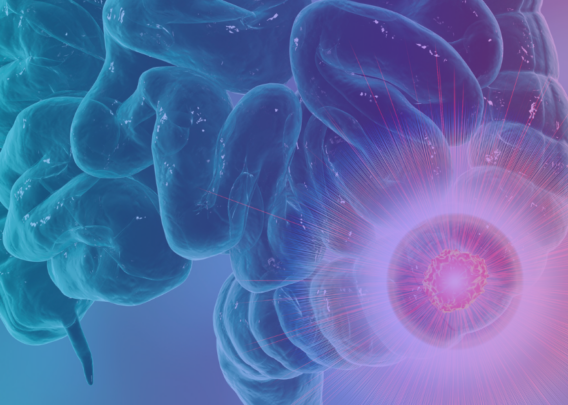
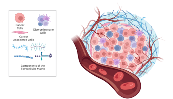
There is growing evidence that the tumor microenvironment (TME) is a crucial modulator of cancer growth, migration, angiogenesis, immunosurveillance evasion, and therapy resistance2. The TME is composed of extracellular matrix (ECM) and various non-malignant cell types, including endothelial, mesenchymal (e.g., fibroblasts and adipocytes) and immune cells (e.g., lymphocytes, monocytes, and neutrophils), all of which interact with tumor cells via secretion of molecules such as growth factors, cytokines, extracellular vesicles, and miRNAs (Figure 1). The ECM in particular has a significant effect on the behavior of cells, as it provides structural and biochemical support. During cancer progression, the stiffness of the ECM influences cells’ proliferation, survival, migration, and differentiation. Thus, understanding the TME is key to understanding overall cancer progression and developing effective therapies.  

Figure 1. The tumor microenvironment

The Need for Better Models 

Animal models have traditionally been considered the gold standard in disease research and drug development, despite the high failure rate of drugs transitioning to the clinic. To improve the translatability of animal models for cancer research, human tumors are often grafted into animals. The xenograft mouse model, for example, is created when the tissue of interest is implanted under the skin of an immunocompromised mouse3,4. The orthotopic mouse model differs in that the human tumor is grafted onto the corresponding organ of interest in the mouse. While these models are more difficult to create, they are more physiologically relevant because the tumors are growing in an organ-specific TME4. Despite using human tumors, however, xenograft and orthotopic mouse models still have limited human relevance, as the immune components and TME are animal-based and do not recapitulate how a tumor progresses in the human body.  

While researchers can create cancer models entirely from human cell sources, they continue to face challenges in fully recapitulating human biological function in a dish. 2D in vitro models are simple and cost-effective models to incorporate into drug screening assays, but their simplicity results in low physiological relevance. Spheroids offer improved 3D cytoarchitecture, but often have issues in uniformity and reproducibility, lack standardized assessments of growth and drug efficacy, and are not high throughput for drug screening5,6.  

Organoids are a powerful alternative to 2D culture that preserves many of the structural and functional traits of their in vivo counterparts, such as a hypoxic microenvironment, cell heterogeneity, ECM interactions, and more in vivo-relevant gene expression patterns7,8. Tumor organoids can either be derived from patient tissue or engineered using induced pluripotent stem cells (iPSCs) and special culture conditions9. Those generated from iPSCs and adult stem cells encompass important tissue features such as architecture, differentiated cell types, and tissue function. Taken together, organoids are comparable to certain in vivo models such as traditional genetically engineered mouse models, cell lines, and patient-derived xenografts (PDX)8. However, organoids lack an organ-specific environment that includes immune components, blood vessels, and different stromal cells. Finally, growth stimulators and inhibitors used in the growth of organoids can affect drug sensitivity, gene expression, and cell signaling pathways3.  

A New Way Forward: Improving 3D Cancer Modeling with Organ-Chips 

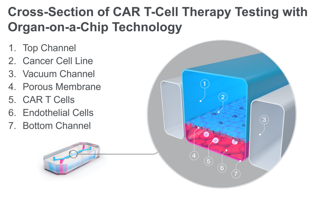
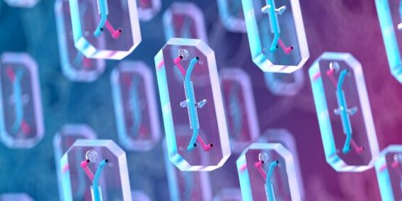
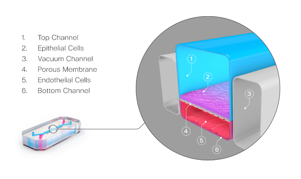
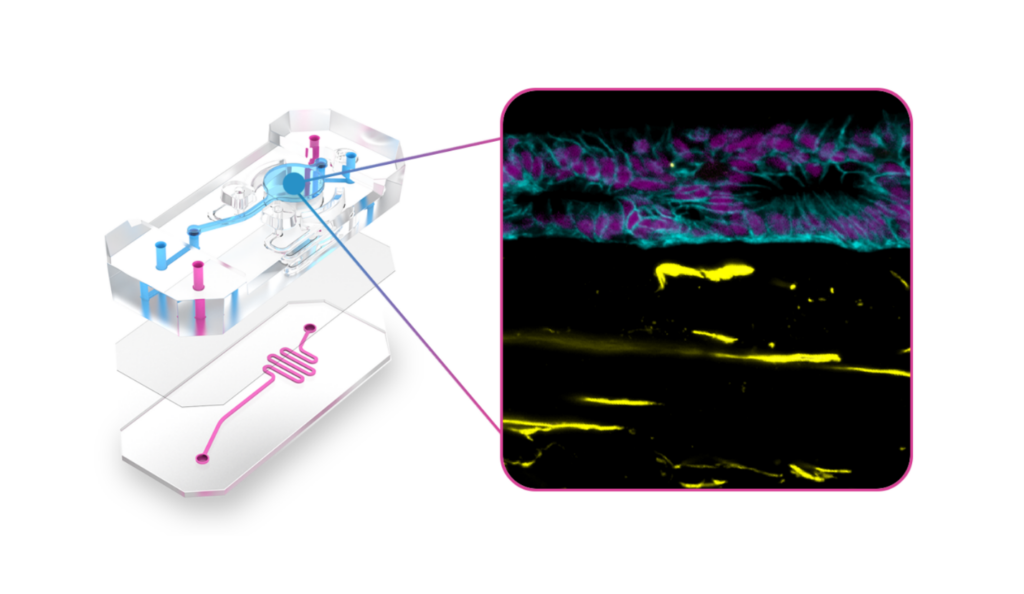
Organ-on-a-Chip technology has been developed to address many of the challenges discussed above. These microfluidic devices emulate in vivo physiology and tissue microenvironments in an organ-specific context by enabling key cellular populations to be added to parallel microfluidic channels that are separated by a porous membrane (Figure 2). Additional features such as biomechanical forces, ECM, and immune cells provide a more human-relevant environment for cells to behave as they would in vivo10–12.

Figure 2. Schematic of an Organ-Chip

Recent advances in the design of Organ-Chip consumables have improved the technology’s ability to recreate the tumor microenvironment. The Emulate Chip-A1 Accessible Chip allows researchers to model complex 3D tissues by incorporating gels up to 3 mm thick within the chip’s accessible culture chamber, integrating stroma into the epithelial layer, creating stratified epithelia, and applying organ-specific biomechanical forces (Figure 3). Further complexity and human relevance can be achieved with Chip-A1 by incorporating circulating immune cells such as PBMCs or CAR T cells.  

Figure 3. Exploded view of Chip-A1 Accessible Chip demonstrating how it has been used to model epithelial-stromal interactions in Barrett’s Esophagus. 

Chip-A1 has already enabled researchers to better understand how the surrounding microenvironment influences cancer progression. In their online article titled ‘Epithelial-Stromal Interactions in Barrett’s Esophagus Modeled in Human Organ Chips,” researchers from the Wyss Institute for Biologically Inspired Engineering at Harvard University used a prototype of Chip-A1 and found that it offered a new approach for studying epithelial-stromal interactions and the broader underlying mechanisms associated with esophageal cancer progression. The team also reported that this model could potentially serve as a tool for personalized drug-response assessments between different patients or genetic subpopulations.   

Conclusion 

Understanding how the tumor microenvironment contributes to cancer progression remains critical for developing more effective therapeutics. Organ-on-a-Chip technology is evolving to meet the in vitro modeling needs for research areas such as cancer, which require enhanced 3D models and more physiologically relevant compound dosing options. Preclinical models that enable researchers to model biology that is more complex, like the TME, will improve clinical translation, and Chip-A1 will give researchers this capability.  

References:

  1. Smietana K, Siatkowski M, Møller M. Trends in clinical success rates. Nat Rev Drug Discov. 2016;15(6):379-380. 
  2. Roma-Rodrigues C, Mendes R, Baptista P V., Fernandes AR. Targeting Tumor Microenvironment for Cancer Therapy. Int J Mol Sci 2019, Vol 20, Page 840. 2019;20(4):840. 
  3. Sajjad H, Imtiaz S, Noor T, Siddiqui YH, Sajjad A, Zia M. Cancer models in preclinical research: A chronicle review of advancement in effective cancer research. Anim Model Exp Med. 2021;4(2):87-103. 
  4. Li Z, Zheng W, Wang H, et al. Application of Animal Models in Cancer Research: Recent Progress and Future Prospects. Cancer Manag Res. 2021;13:2455. 
  5. Pinto B, Henriques AC, Silva PMA, Bousbaa H. Three-Dimensional Spheroids as In Vitro Preclinical Models for Cancer Research. Pharmaceutics. 2020;12(12):1-38. 
  6. Han SJ, Kwon S, Kim KS. Challenges of applying multicellular tumor spheroids in preclinical phase. Cancer Cell Int 2021 211. 2021;21(1):1-19. 
  7. Salinas-Vera YM, Valdés J, Pérez-Navarro Y, et al. Three-Dimensional 3D Culture Models in Gynecological and Breast Cancer Research. Front Oncol. 2022;12:826113. 
  8. Tuveson D, Clevers H. Cancer modeling meets human organoid technology. Science (80- ). 2019;364(6444):952-955. 
  9. Fan H, Demirci U, Chen P. Emerging organoid models: Leaping forward in cancer research. J Hematol Oncol. 2019;12(1):1-10. 
  10. Huh D, Matthews BD, Mammoto A, Montoya-Zavala M, Hsin HY, Ingber DE. Reconstituting Organ-Level Lung Functions on a Chip. Science (80- ). 2010;328(5986):1662-1668. 
  11. Kasendra M, Luc R, Yin J, et al. Duodenum intestine-chip for preclinical drug assessment in a human relevant model. Elife. 2020;9. 
  12. Kerns SJ, Belgur C, Petropolis D, et al. Human immunocompetent Organ-on-Chip platforms allow safety profiling of tumor-targeted T-cell bispecific antibodies. Elife. 2021;10. 

Solid Tumors: The Next Frontier of Cancer Immunotherapy

In the last decade, the realm of cancer therapy has taken groundbreaking strides, with Chimeric Antigen Receptor (CAR) T-cell therapy being one of the most promising new types of treatment. Having shown potential in treating blood cancers, CAR T-cell therapies are now setting their sights on a more complex adversary: solid tumors. This article explains what CAR T-cell therapy is, why solid tumors are such a challenging target, and how researchers can use advanced in vitro models to gain a more human-relevant understanding of CAR T efficacy. 

Understanding CAR T-Cell Therapy 

At its core, autologous CAR T-cell therapy is a form of immunotherapy where a patient’s own immune cells are modified so they recognize and kill cancer cells more effectively. This is done by adding a gene for a receptor, called a chimeric antigen receptor (CAR), which is designed to identify a specific tumor antigen. These modified cells are infused back into the patient, where they hunt down and kill the cancer cells. 

To date, researchers have successfully developed six FDA-approved CAR T-cell therapies for blood cancers, including leukemia, lymphoma, and multiple myeloma. This has proved invaluable for those in difficult-to-treat patient populations who exhibit little to no response to conventional therapies. 

However, these hematological malignancies only represent a small portion of all cancer cases and deaths—about 90% of cancers are solid tumors. Unfortunately, efforts to adapt CAR T-cell therapy for solid tumors have, to date, proved challenging. 

Stay Up to Date with the Emulate newsletter

The Challenges of CAR T for Solid Tumors 

High-mortality, treatment-resistant solid tumors—including lung, breast, colorectal, and pancreatic cancer—are prime candidates for immunotherapy. However, the relative complexity of the heterogeneous solid tumor environment creates novel immunological obstacles that are not seen in blood cancers. 

The challenges are many, but a few highlights include: 

  • Selective Trafficking and Migration: For blood cancers, immunotherapies are administered in the same location where the cancerous cells reside—the circulatory system. But for solid tumors, this is just the start of the journey. After being administered into the blood stream, the immune cells must selectively traffic out of the vasculature at the site of the tumor and then migrate to the tumor tissue. This is a challenging but critical journey, as the CAR T cells must reach the tumor in adequate numbers to effectively kill the cancerous cells, while avoid attacking healthy tissue.  
  • Tumor antigen heterogeneity: In hematological tumors, antigen expression is relatively stable and uniform. But in sharp contrast, solid tumors can exhibit significant variability in antigen expression, both within a single tumor and between patients. This means that CAR T-cell therapy targeting a specific antigen might only attack certain parts of the tumor, leaving other parts untouched. It may also present the need for a more personalized or multi-antigen-targeted CAR T approach. 
  • Tumor Microenvironment (TME): The area surrounding solid tumors is a unique cellular and chemical landscape, where inhibitory immune cells, immune checkpoints, and cytokines create an environment that is naturally hostile to immune cells, including CAR T-cell therapy. This means that once CAR T cells reach the tumor, they can quickly become suppressed or inactivated by immunosuppressive factors in the TME, hindering their ability to effectively destroy cancer cells. 

The Need for Human-Relevant Research Models 

While enthusiasm for solid tumor CAR T-cell therapy is strong, FDA-approved therapies have so far remained out of reach largely due to the challenges described above. A major limiting factor in overcoming these challenges has been a lack of preclinical research models that can adequately recreate the complex journey that CAR T cells must undertake in the human body to be effective. The models that researchers rely on for developing immunotherapy fall into two main categories: in vitro models and animal models. 

2D cell models often include tumor cell lines or patient-derived organoids (PDO) cultured in a static dish, where the CAR T cells are be administered directly to the tumor cells to evaluate killing efficacy. However, these models only allow researchers to evaluate the end of the CAR T cell journey and completely ignore the aspect of CAR T cell tracking and migration. 

Animal models remain the cornerstone of preclinical immunotherapy testing and have provided important contributions to hematological cancer CAR T-cell therapies that are on the market today. One example is patient-derived xenografts (PDX) models, in which human patient-derived tumors are implanted in immunodeficient mice. However, while animal models enable a more complete understanding of immunotherapy effect—allowing the CAR T cells to be intravenously administered and traffic to the tumor as they would in humans—they suffer from large species differences in responses and relatively poor insights into mechanism of action, limiting their successful translation to human clinical response. 

To truly understand the dynamics of cancer immunotherapy for solid tumors and develop effective treatments, researchers need more advanced models that can capture the complex intricacies of human biology and immune response. 

A Path Forward for Solid Tumor Immunotherapy Development 

For CAR T-cell therapy’s potential to be fully realized in solid tumors, it is vital that its therapeutic candidates’ behavior, efficacy, and challenges be studied in a context that is as close to human as possible. 

Fortunately, Organ-on-a-Chip technology can offer a path forward. Organ-Chips are microengineered devices that emulate the architecture, functionality, and physiological responses of vascularized human organs. They provide a dynamic platform that closely replicates human tissue-tissue interfaces, fluid flow, and mechanical forces, creating an environment where CAR T cells can be studied in a setting that more accurately mirrors the human body.  

Recently, this technology has been used to study the efficacy of CAR T-cell therapy in a model of non-small cell lung carcinoma (NSCLC). By co-culturing an NSCLC cell line and lung-specific vascular cells, researchers were able to model the journey that CAR T cells undergo in vivo. After administering the immunotherapy in the chip vasculature—just like its intravenous administration in the body—researchers saw how the CAR T cells attached to the vasculature, migrated to the tumor cell-containing channel, and subsequently exhibited antigen-dependent killing of cancer cells. Through imaging and effluent analysis, they could even quantify the amount of migration and killing as well as the levels of exhaustion markers for CAR T cells. A common treatment approach of administering a co-therapeutic alongside CAR T cells was even modeled on-chip, where it demonstrated improved CAR T-cell migration to the site of the solid tumor cell line. As this technology becomes more widespread, it may enable immunotherapy companies to accelerate the successful development of the first solid tumor CAR T-cell therapy. 

Conclusion 

Solid tumors continue to present numerous challenges for CAR T-cell therapy researchers, including efficient CAR T cell trafficking, migration, and infiltration, as well as overcoming the hostile tumor microenvironment. 

As researchers look to surmount these obstacles, advanced cell culture technologies like Organ-Chips may provide a more human-relevant and predictive assessment of CAR T cell efficacy, improving translation from pre-clinical testing to clinical success. With a number of CAR T-cell therapies in development around the world, the horizon seems promising, providing hope that researchers may be able to widen the reach of CAR T cells beyond blood cancer and into the more formidable realm of solid tumors.


Cancer immunotherapy approaches are having their moment in the clinical spotlight. Over two decades of work characterizing potent activators of the immune system have delivered impressive results with biologic drugs like atezolizumab (Tecentriq) and cell therapies like chimeric antigen receptor T-cell (CAR-T) treatment. 

An illustration of antibody molecular structure
Antibody Molecular Structure

T-cell engaging bispecific antibodies (TCBs) are an emerging class of engineered immunotherapeutic agents that simultaneously bind a cancer cell antigen and the T-cell CD3 receptor. This mechanism activates the T-cell while physically linking it via the target antigen to the cancer cell, which is useful for cells that do not express high levels of immunogenic proteins on their surface or cancers that evade the immune response. As a promising oncotherapy, TCBs are being explored clinically for solid tumor and blood cancers. However, the target antigens are rarely tumor-exclusive; they may be expressed in normal tissues leading to on-target/off-tumor adverse effects that compromise patient safety. 

Reliable TCB safety evaluations are critically important to ensuring that immunotherapies are both effective and well-tolerated. The ability to accurately predict therapeutic efficacy and safety remains a challenge. Animal models and two-dimensional (2D) cell culture models of tissues and organs, such as transwells, have been used for the purpose with some success, but are also subject to limitations. Many therapeutic drugs are designed to target human biology, which differs from the biology of animals. 2D cell culture models lack circulation, immune cells, and the proper microenvironment for target expression and immune cell activation. For example, standard in vitro models also do not capture air interfaces or mechanical forces that mimic breathing and peristalsis critical for the functional properties of organ tissues. Animal preclinical models are often not informative for predicting human cancer immunotherapy-mediated adverse events due to lack of cross-reactivity with drugs targeting human-specific epitopes. 

Organs-on-Chips aim to overcome these limitations by modeling the complex multifactorial microenvironment of native tissues and organs and more accurately reflecting the complexity and specificity of human immunity. In a study published in eLife and available on PubMed, researchers from Roche and Emulate assessed in vivo target expression and toxicity of TCBs targeting folate receptor 1 (FOLR1) or carcinoembryonic antigen (CEA) to design and validate Organs-on-Chips as a more human-relevant platform to determine safety and efficacy. 

  • Research Area: Immunotherapeutic drugs, toxicity testing
  • Organisms: human
  • Sample Types: human Lung Alveolus-Chip, human Duodenum Intestine-Chip, human Colon Intestine-Chip
  • Research Question: Can organs-on-chips be used as human organ models to evaluate the on-target/off-tumor safety and efficacy profiles of TCBs?


Experimental Overview


Lung Toxicity targeting folate receptor-1 (FOLR1)

A TCB to folate receptor-1 (FOLR1), overexpressed in ovarian, lung and breast cancers as well as in normal tissue, was chosen for study of on-target/off-tissue toxicity. This TCB showed high efficacy in human breast tissue xenografts but resulted in severe on-target lung immune toxicity in monkeys.

To evaluate the safety of FOLR1 TCBs, an Alveolus Lung-Chip model was developed consisting of primary lung alveolar epithelial cells in the upper chamber exposed to air, with primary microvascular cells established in the lower chamber together with peripheral blood mononuclear cells. Immune activation was measured by T-cell crosslinks to the FOLR1 expressing target cells mediated by the TCB, subsequent T-cell activation, cytotoxic granule release and target cell apoptosis.

In order to determine if cellular immune responses were different depending on the affinity of the antibody for its tumor target, two TCBs with different affinities for the target molecule were tested. FOLR1(hi)-TCB with a high affinity for FOLR1, and FOLR(lo)-TCB with a lower affinity for the same target receptor were examined for cell killing efficacy and for off-target cytotoxicity. The authors found that the lower affinity molecule had a better safety profile than the higher affinity one, which was also recapitulated in a primate animal model. Notably, a transwell culture version of this experimental setup did not accurately reproduce the animal results.


Gastrointestinal toxicity targeting carcinoembryonic antigen (CEA)

Carcinoembryonic antigen (CEA) is a cell surface antigen expressed in varying amounts in the gut and is often overexpressed in colorectal cancers. A second study with CEA-engaging TCBs, which are specific for humans and not cross-reactive with mouse or monkey CEA, was done using Organ-Chips to assess potential intestinal toxicities.

The researchers profiled two types of Organ-Chips (Duodenum-Intestine and Colon-Intestine) both demonstrating tight, polarized epithelial barriers and mature enterocytes to assess the safety of high- and low-affinity CEA TCB variants. The researchers observed physiologically relevant expression of the CEA target within these intestinal models, and assessed immune cell crosslinking to the target antigen, activation and epithelial cell death.

They saw that the Colon Intestine-Chip had higher expression of the CEA target than the Duodenum Intestine-Chip on the cell surfaces. The Colon Intestine-Chip reproduced the distribution and density found in the native tissue, which was not replicated in static transwell culture. Furthermore, the Intestine-Chip predicted CEA-mediated on-target toxicity, the extent of which was governed by antibody affinity and target abundance, with the colon being more liable to damage compared with the duodenum.

The authors also concluded that the high affinity TCB might induce off-tumor toxicity even in tissues with low target expression such as in the small intestine.


Conclusions

Human immunocompetent models of the lung and intestine were created using Emulate’s Organ-on-a-Chip technology, and they were validated for profiling lung and intestine toxicity of engineered TCBs.

  • The Alveolus Lung-Chip reproduced FOLR1 TCB-mediated lung toxicity and instructed the design of a lower affinity antibody with a more favorable safety profile, verified in non-human primates.
  • The Colon Intestine-Chip models highlighted the safety liabilities of a TCB that targets a human antigen that lacks an animal counterpart. These experiments showed that the Organs-on-Chips could show sensitivity to antibody affinity and show differences in target-dependent toxicities in different regions of the intestine.

The Organs-on-Chip models developed in this study demonstrate clear advantages over traditional in vitro efficacy and safety models. “With functional features of the human immune system, these models represent a promising alternative platform on which to perform immunotherapeutic safety testing, format selection and optimization” said Lauriane Cabon of Roche, a lead author on the study. In the hands of academic and pharmaceutical researchers, Organ-Chips will reduce reliance on animal-based safety assessments, inform antibody format selection, shed light into the mechanistic underpinnings of toxicities, and potentially support identification of new clinical cancer biomarkers.

“Engineered immunotherapies are highly efficacious but have shown significant clinical safety liability sometimes missed in conventional safety profiling. Advanced immunocompetent Organ-Chip models with the ability to predict off-tumor cytotoxicity in multiple affected tissue types could have a real impact on the drugs selected for clinical assessment, reduce the use of animals, and improve patient outcomes” said S. Jordan Kerns, a lead co-author on the study.