Writing in a recent issue of Nature Reviews Drug Discovery, Jack Scannell, author of Eroom’s law, and a team of experts stated plainly that predictive validity is “the thing that nearly everyone already believes is important [but which] is more important than nearly everyone already believes.” In other words, the drug development industry knows that predictive validity is important, but Scannell and his co-authors argue that it is far more important than most understand it to be.  

Predictive validity describes a tool’s ability to reliably predict a future outcome. The drug development industry has struggled for centuries to find preclinical models that accurately predict which drug candidates will be both safe and effective in humans. Despite remarkable advances in science over the past half-century, including the advent of tools like next-generation sequencing and CRISPR editing technology, success in drug development remains rare. 

“Successful drug discovery is like finding oases of safety and efficacy in chemical and biological deserts,” writes Scannell and his co-authors, referring to the 90% to 97% clinical trial failure rate that the industry is familiar with. They argue that the drug developers have spent recent decades attempting to improve success rates through brute force scale—the equivalent of searching for oases by simply running over more desert terrain. Instead, they argue, the industry should focus on improving the predictive validity of existing preclinical models, as these are the compasses that will lead to success.  

The Faulty Preclinical Models Still In Use 

At every stage of the drug discovery and development process, researchers must choose which compounds are worth investing in and which should be left by the wayside. To make these difficult decisions, they rely on tools such as cell lines, animal models, or computer modeling to predict how these prospective compounds will behave in patients. Naturally, the quality of these tools—specifically their predictive validity—has a direct impact on the quality of drug development decisions.  

If a model has perfect predictive validity, you can trust that a drug’s behavior in the model will perfectly match its behavior in patients. However, researchers have yet to find such a model—a reality that has greatly limited drug development success.  

Take for instance the use of rodents to study ischemic stroke. Such models are easy to use and widely available, making them particularly attractive for drug testing. And they are robust—that is, the performance of compounds in one rodent model is likely to replicate in another.  

However, there are important genetic and physiological differences between rodents and humans that severely reduce these models’ predictive validity for drug performance in humans. Not surprisingly, these rodent models select drugs that are both safe and effective for rodents, but not necessarily for humans. Scannell and his co-authors underscore this point by saying that “it now seems likely that non-diabetic normotensive rodents respond better to a range of neuroprotective drugs than do the bulk of elderly human patients with stroke.” 

In other words, a rat, no matter how fancy, can only go so far in mimicking human physiology and disease response. In reality, no model can be perfectly predictive. What they can do is provide narrow ranges of value—what Scannell and his co-authors refer to as “domains of validity.” 

Stay Up to Date with the Emulate newsletter

Domain of Validity and the Limitations of Models 

A domain of validity is the specific context in which a model is most predictive. Take tumor cell lines in oncology, for example. For nearly a century, researchers have been developing cytotoxic drugs to treat cancer based on studies in which human cancer cell lines are grown as 2D monolayers. These cells are typically fast growing, genetically homogenous, and able to be rapidly scaled up for high-throughput screening. However, their fast-growing nature leaves them uniquely susceptible to cytotoxic drugs—something researchers have known since the late 1960s1.  

Many cancers, though, comprise heterogeneous and slow-growing cells. It’s no surprise then that between 2000 and 2015, oncology had the highest clinical trial failure rate of any major therapy area at 97%2,3. Human cancer cell lines are valuable tools, but they are representations of the small subset of fast-growing cancers.  

Put another way, many cancer cell lines are at their peak predictive validity only in the narrow domain of predicting drug effects in fast-growing, homogenous tumors. By using these cells to represent tumors broadly, researchers unknowingly extended the model well beyond its narrow domain of authority and, as a result, overestimated how effective cytotoxic drugs would be in tumors containing slow-growing cells.  

These examples show that it is critical for researchers to carefully consider their models’ predictive limitations when making drug development decisions. They also exemplify that the standard models for preclinical decision-making—namely animals and conventional cell culture—are severely limited, and this has greatly curtailed the pace of drug development and driven the cost of bringing a new drug to market above $2 billion.  

Many have tried to compensate for models’ predictive limitations by increasing model throughput, running the same models at a higher pace to gather more data. However, Scannell and his co-authors urge the industry to focus instead on developing better, more predictive models.    

Towards Better Model Predictive Validity 

In previous papers, Scannell and many of his co-authors have argued that incremental improvements in the predictive validity of a model can have a far greater impact on drug development success than simply improving the number of compounds being screened4,5. Clearly, part of improving predictive validity requires researchers to evaluate their models and establish domains of validity for each.  

Unfortunately, doing so is a complex process that often lacks incentives. It may take years or decades to determine if a tool is truly predictive of clinical outcome, and performing such an analysis would require dedicated funds and clear definitions of what qualifies as sufficiently predictive.  

Fortunately, Scannell and his co-authors offer several suggestions for how we can improve model validity. An abbreviated list is presented below: 

  • Evaluate the models we use as best we can. This should include the collection of structured and consistent data on model performance and an explicit effort to both reduce bias and ensure that criteria for evaluation are informed by expert opinions. 
  • Fund retrospective studies that compare tools to determine why some models are successful and others are not. 
  • Establish a system for institutional learning such that, after evaluations are performed, others can learn from it and iterate. 
  • Foster inter-organizational information sharing of detailed protocols, calibration methods, calibration results, decision tools’ contexts-of-use, and associated craft skills.  

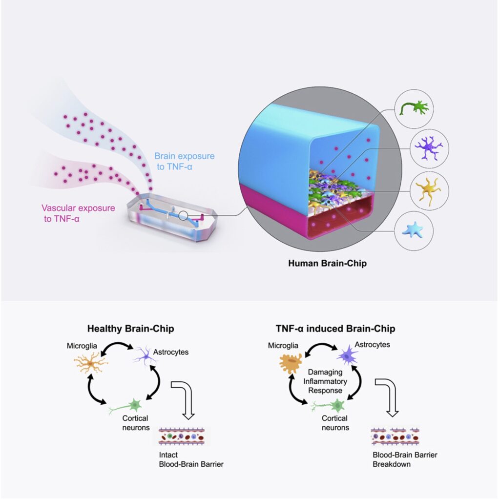
Improving Drug Development with Organ-Chips 

Ultimately, these authors make it clear that the drug development industry—as well as the patients who rely on it—stand to benefit greatly from close examination of models’ predictive validity. Ideally, drug development pipelines would not be static or adherent to models based on tradition, but rather would select the models based on superior and complementary domains of authority.  

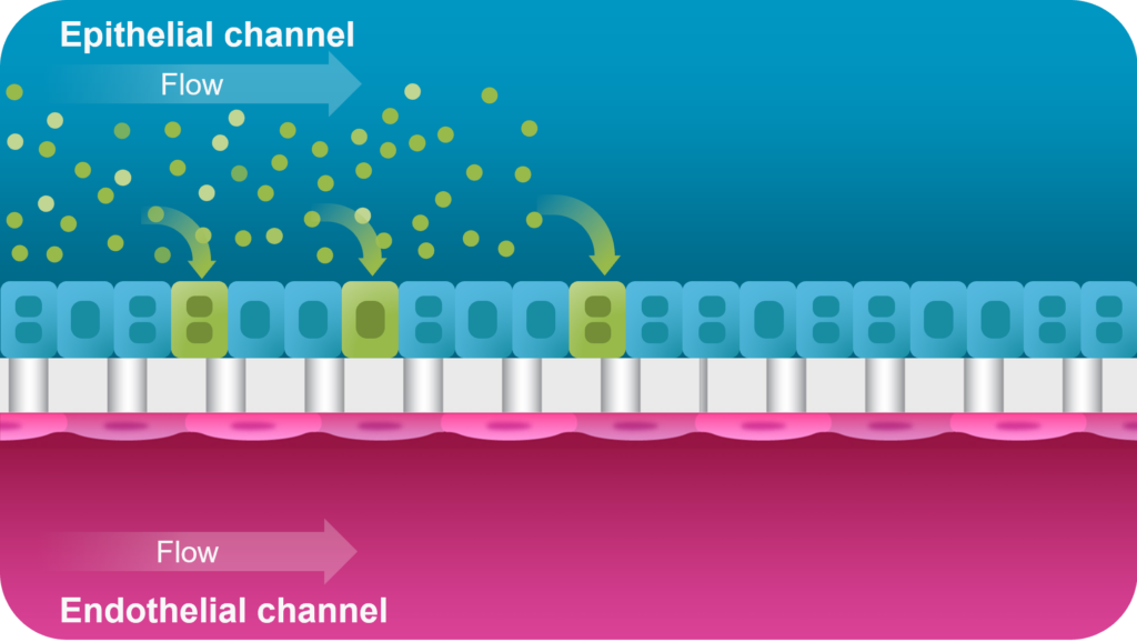
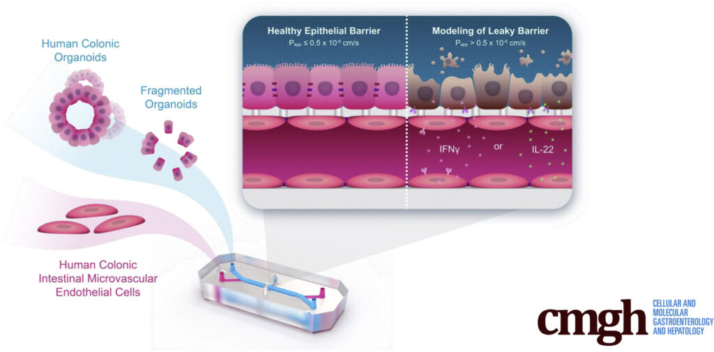
This is where Organ-on-a-Chip technology can make a big impact. Organ-Chips are unique model systems that integrate living human cells, tissue-tissue interactions, tissue-specific proteins, chemical environments, and biomechanical forces to emulate human tissues. Several lines of evidence show that Organ-Chips can closely resemble in vivo human organ behavior and may be beneficial for predicting drug toxicity, among many other applications.  

Exemplifying this, a recent paper in Communications Medicine, part of Nature Portfolio, showed that the Emulate human Liver-Chip model was far more predictive of drug-induced liver toxicity relative to animal and spheroid models. In collaboration with Scannell, the team behind the study then showed that integrating the Emulate human Liver-Chip into drug development pipelines could result in more than $3 billion dollars in excess productivity for the drug development industry, owing to the model’s improved predictive validity. 

Along with the Emulate human Liver-Chip study, the argument made by Scannell and his co-authors in Nature Reviews Drug Discovery may be a critical moment of reckoning for drug developers. Now is the time to prioritize predictive validity and begin integrating better, more human-relevant tools into drug development programs.  

References 

  1. Skipper HE. The effects of chemotherapy on the kinetics of leukemic cell behavior. Cancer Res. 1965 Oct;25(9):1544-50. PMID: 5861078. 
  1. Chabner, B., Roberts, T. Chemotherapy and the war on cancer. Nat Rev Cancer 5, 65–72 (2005). https://doi.org/10.1038/nrc1529 
  1. DiMasi, J.A. (2001), Risks in new drug development: Approval success rates for investigational drugs. Clinical Pharmacology & Therapeutics, 69: 297-307. https://doi.org/10.1067/mcp.2001.115446 
  1. Scannell JW, Bosley J (2016) When Quality Beats Quantity: Decision Theory, Drug Discovery, and the Reproducibility Crisis. PLoS ONE 11(2): e0147215. https://doi.org/10.1371/journal.pone.0147215 
  1. Ringel, M. S., Scannell, J. W., Baedeker, M., & Schulze, U. (2020, April 16). Breaking eroom’s law. Nature News. Retrieved March 21, 2023, from https://www.nature.com/articles/d41573-020-00059-3 

Gene therapies have generated both excitement and hope, as they have the potential to correct underlying genetic defects and cure diseases. However, one of the major hurdles gene therapy researchers face is delivering enough molecular material into target cells to make a difference—a key aspect of this type of treatment. 

Many labs have turned to adeno-associated viruses (AAVs), which can be excellent vehicles for delivering genetic material and/or editing tools. They persist in host cells and present low immunogenicity and toxicity risks. In addition, AAVs are easily modifiable, stable, and adaptable to many tissues. However, the same labs have faced difficulty when researching AAVs using conventional models, such as 2D cell cultures and animals. 

The Emulate Liver-Chip offers a more human-relevant way of testing viral vectors during early development and preclinical studies compared to conventional models. The Liver-Chip incorporates multiple cell types in a dynamic microenvironment, allowing researchers to assess virus-mediated transduction efficiency and toxicity to develop safe, effective treatments. 

BARRIERS TO GENE THERAPY

AAVs have been used in therapies for hemophilia, inherited liver diseases, neurological conditions, and other disorders. While there are only a few gene therapies in the clinic, there are many on the horizon.  

But the distance to that horizon has become problematic, as AAV-based therapy development has been slow and costly. Since labs lack good pre-clinical models to robustly study toxicity and gene delivery efficacy, they are being forced to take the long road. 

A major concern for any emerging gene therapy is hepatotoxicity risk, as even the most precisely targeted vectors often accumulate in the liver. In fact, hepatotoxicity is the most common stumbling block seen in clinical trials for AAV therapeutics.  

Current pre-clinical studies largely use 2D cell cultures and animal models. In vitro cell models provide inadequate data because they lack the complexity to capture in vivo physiology. These 2D models generally have one cell type—often hepatocytes for liver studies—but do not incorporate fluid flow and other biomechanical forces found in vivo. In addition, non-human primate studies are slow, costly, and tightly regulated, limiting the pace of viral vector optimization.   

The life sciences industry needs advanced technologies that accurately mimic human physiology to effectively translate pre-clinical results into successful human trials. Because Organ-on-a-Chip technology more closely recapitulates in vivo functions, it predicts human responses far more reliably than other models. Organ-Chips accurately replicate human physiology by including dynamic media flow, cellular complexity, 3D cellular architecture, and cell-cell signaling. 

By providing a more human-relevant in vitro platform for AAV-based therapeutic development, Organ-Chips can help researchers rapidly iterate on AAV designs to accelerate gene therapy development, reduce clinical trial attrition, and bring new products to patients faster. 

THE EMULATE LIVER-CHIP IN ACTION

Cross-section of the Emulate Liver-Chip

The Liver-Chip co-cultures primary human hepatocytes and liver sinusoidal endothelial cells under tissue-specific media flow. It also enables the addition of Kupffer and stellate cells, allowing researchers to capture additional cell-cell interactions. As a result, it better represents human in vivo liver tissue than conventional approaches and facilitates measurements of AAV transduction efficiency and toxicity differences across space, time, and cell types.  

To assess the Emulate Liver-Chip’s ability to predict AAV transduction efficiency and hepatotoxicity risk, chips were treated with six conditions—including multiple MOIs (multiciplicities of infeciton) of AAV2 and AAV6 vectors—administered for 24 hours via the epithelial channel. After treatment, they were monitored and measured for transduction efficiency and safety. 

During the seven-day monitoring period, the chips displayed concentration-, time-, and serotype-dependent transduction. Researchers also assessed morphology and functional liver endpoints (albumin, ALT) and confirmed that there were no toxic responses, showing how the model can be used to assess AAV serotypes’ potential to induce toxicity in human liver tissue. 

Liver AAV Schematic
AAV Transduction in the Liver-Chip

A proof-of-concept study showed that the Emulate Liver-Chip can model AAV vector transport across vascular tissue for subsequent transduction of epithelial tissue, allowing researchers to emulate the intravenous administration route for many AAV-based gene therapies. Furthermore, AAV was administered via the vascular channel and demonstrated successful hepatocyte transduction. 

The Emulate Liver-Chip successfully demonstrated its potential to accelerate workflows for AAV gene therapy development and preclinical testing. With this important tool, researchers can accumulate actionable data in weeks, rather than the months these studies might take in animal models. Since the Liver-Chip facilitates multiple rapid iterations—which is critical to designing effective gene therapies—scientists can gain deeper insights into AAV design to bring safe, effective treatments—even potential cures—to patients. 


Inflammatory bowel disease (IBD) is increasing around the world. In 1990, around 3.7 million people had the condition; by 2017, that number had increased to 6.8 million. Nearly half of IBD patients don’t respond to current treatments, and even for the lucky ones, therapeutic efficacy can wane over time. As a result, there is an urgent need to develop a new generation of IBD therapies. 

Unfortunately, ineffective drug development models are hampering the search for more effective treatments. Conventional two-dimensional (2D) cell models only capture bits and pieces of IBD’s complexity, and many three-dimensional (3D) culture models like organoids fall short because they lack critical biological features, such as vasculature and biomechanical forces. 

Animal models have their own drawbacks, as their immune systems fail to replicate many of the mechanisms associated with human immunity. 

“If you look at the physiology of cardiac muscle or neurons between humans and mice, they’re fairly similar,” said Christopher Carman, PhD, Director of Immunology at Emulate. “There’s more divergence in immunology, and it can be really challenging to extract meaningful insights around immune-system-driven mechanisms. That’s why so many therapeutics fail.” 

To remedy this, Emulate has developed a Colon Intestine-Chip that combines primary human tissue, vasculature, mechanical forces, and (most importantly) immune cell recruitment to recapitulate the biology that drives IBD.  

Understanding How IBD Evolves 

IBD begins with an unknown tissue insult, and the body responds by producing inflammatory cytokines and chemokines. In turn, these proteins recruit immune cells to the intestine, inducing further inflammation. 

This process generates a cytokine cascade. Two proteins in particular, interferon gamma (IFNγ) and IL-22, act directly on colon epithelial cells, driving cell death, microvilli loss, and destruction of the tight junctions that guard intestinal permeability. 

“That is a critical hallmark of this disease,” said Carman. “As a result, intestinal material, including bacteria and bacterial products, leak into the interstitial space, driving even more inflammation.” 

Making the Colon Intestine-Chip 

A cross-section of the Colon Intestine-Chip. The parts are diagrammed.
Cross Section of the Colon Intestine-Chip

The Emulate Colon Intestine-Chip was designed to precisely recapitulate this inflammatory cascade.  

This advanced, in vitro intestine model incorporates primary human biopsy tissue cultured into organoids. Critically, the cells retain their “stemness,” meaning they replicate the stem cell niches that are constantly regenerating in human intestines.  

After the organoids are dissociated, they are seeded in the top channel of the Organ-Chip. The bottom channel contains primary human intestine-derived microvascular endothelial cells, which are in close proximity to the epithelial cells, as they would be in vivo. The channels are separated by a porous membrane coated with tissue-relevant extracellular matrix proteins. 

From there, mechanical forces on the chip—physiologic flow and cyclic stretch—replicate intestinal peristalsis, which improves cell morphology and functionality while supporting more accurate gene expression. 

As a result, epithelial tissues respond to microvasculature cues, and the epithelial cells differentiate into all three major epithelial types at the appropriate ratios.  

With this, the Emulate Colon Intestine-Chip is able to model IBD from the initial insult to the cytokine cascade, demonstrating along the way selective immune cell recruitment, cell death, and tight junction loss. This model can be applied to study inflammation-specific immune recruitment from vasculature into epithelial tissue and subsequent downstream impacts. 

“We have shown that this Organ-Chip strongly reflects what we see in primary human tissue,” said Carman. “It develops proper tight junctions and a strong functional barrier. On the molecular level, we see transcriptional signatures that are highly reflective of primary human tissue.”  

This model has demonstrated the efficacy of small molecule inhibitors that target IFNγ and IL-22 signaling pathways, meaning researchers can use it to validate  clinically relevant drug candidates designed to prevent barrier dysfunction. 

Illustration of Immune Cell Recruitment in the Colon Intestine-Chip

Selectively Generating Inflammation 

One of the Organ-Chip’s most important abilities is the selective recruitment of immune cells. This selectivity comes from tissue-specific adhesion molecules on both endothelial and immune cells, which must be highly specific to bind.   

Around 30% of the body’s circulating immune cells are customized for work in the intestines. They have a molecule called α4β7 integrin that binds to an endothelial molecule called MAdCAM-1, which is preferentially expressed in the colon endothelium and upregulated in response to inflammatory cues. 

One of the major ways the Colon Intestine-Chip replicates IBD biology is by expressing MAdCAM-1 in response to inflammatory stimuli, giving it tremendous relevance for therapeutic discovery. 

“The α4β7 integrin/MAdCAM-1 adhesion molecule axis is an important therapeutic target,” said Carman. “If we can interfere with that adhesion, we can potentially interrupt the inflammatory cascade. And because this mechanism is selective to the gut, any therapeutic that targets these adhesion molecules would be highly specific to the intestinal system.” 

Image of the Application Note "Modeling Inflammation-Specific Immune Cell Recruitment in the Colon Intestine-Chip"
Download the application note to see the data.

“One drug, AJM300, is in phase three clinical trials right now and is showing promising safety and efficacy,” said Carman. “We validated that efficacy in our model. We also used the model to study the corticosteroid dexamethasone, which has been a mainstay in IBD treatment for many years. We recently published the data in an application note.” 

The Colon Intestine-Chip provides a more complete picture of human IBD pathogenesis, delivering a human-relevant platform to test drug efficacy. However, for Emulate, it’s just the beginning. Inflammation plays a major role in many conditions, and creating models that effectively replicate those pathways will be essential in validating and advancing therapeutic compounds to support better care. 

“This IBD model is our first foray into inflammation,” said Carman. “We’re planning on developing many variations on this theme to create better tools for a variety of inflammation-driven indications.”   

For more information on Emulate’s IBD model, please download Modeling Inflammation-Specific Immune Cell Recruitment in the Colon Intestine-Chip. 

A summary of the paper published in iScience: A Microengineered Brain-Chip to Model Neuroinflammation in Humans

A growing and persistent storm of inflammatory signaling often accompanies diseases like Alzheimer’s, Parkinson’s, and Huntington’s—neurodegenerative diseases that affect more than a billion people worldwide. Decades of research have shined a light on the complex interplay among cell types of the brain that leads to the propagation of neuroinflammation. Yet, in spite of substantial advances in our understanding of the biology underlying neurodegenerative diseases, development of therapeutics that treat these conditions has not kept pace.

One potential reason for this discordance is the lack of human-relevant models on which to test new therapeutics. Because of their artificiality, conventional, two-dimensional cell cultures fail to capture the complexity of the brain neurovascular unit and the many cell-cell interactions that mediate a drug’s effects. Additionally, myriad differences between species in neurobiology limit the physiological relevance of animal models.

In a recent study published in iScience, Emulate researchers describe a new model of neuroinflammation based on Organ-on-a-Chip technology. By co-culturing multiple human brain cell types in a three-dimensional microphysiological system, the authors were able to more accurately recreate the structure and function of the human neurovascular unit and present a physiological model of neuroinflammation with significant advantages over contemporary models. Such a model may prove to be a critical tool in the development of novel therapeutics for neurodegenerative conditions.

Experimental Overview

Research Area: Brain; mechanisms of neuroinflammation in neurodegenerative diseases

Organisms: Human

Sample Types: Brain-Chip

Research Question: Can a human Brain-Chip accurately model human brain neuroinflammation?

Results:

  • Microengineered human Brain-Chips develop mature excitatory and inhibitory cortical neurons juxtaposed with glia cells, astrocytes, pericytes, and a tight layer of endothelial-like cells; collectively, these mimic protein expression, permeability, and architecture of the in vivo human neurovascular unit (NVU).
  • Relative to conventional Transwell cell culture systems, the Brain-Chip shows an RNA expression profile that more closely resembles adult human cortex tissue (a first for microphysiological systems).
  • Perfusion of the Brain-Chip with TNF-α elicits key inflammatory features seen in neurodegenerative disease patients, such as glial activation, the release of proinflammatory cytokines, and increased barrier permeability.

Conclusion: Collectively, results from this paper indicate that the Emulate Brain-Chip more closely mimics human cortical tissue relative to alternative models. As such, this “brain-on-a-chip” model may help researchers better understand the biology of human cortical tissue and be used as a tool to help elucidate how either pathogenic signaling or therapeutics influence cortical tissue health. 

Emulating the Neurovascular Unit

Neurodegeneration is driven, at least in part, by the establishment of a proinflammatory microenvironment. Compelling evidence suggests that local stress or systemic inflammation can compromise the blood brain barrier, enabling the entry of pro-inflammatory cytokines into the neurovascular unit and subsequent neuroinflammation. Additionally, reactive astrocytes and activated microglia are strongly implicated in blood-brain barrier breakdown and in the establishment of a neurodegenerative microenvironment.

The complex interactions of each cell type within the neurovascular unit appear to be critically important in driving disease progression and are also exceedingly difficult to model. Contemporary in vitro models range from two-dimensional co-cultures to three-dimensional multicellular organoids. However, no system has been able to simultaneously replicate neurovascular structure, function, and cell heterogeneity—all important factors that may affect cell behavior and therapeutic effectiveness.

To address this challenge, Emulate researchers are developing the Brain-Chip, which contains human primary astrocytes, primary pericytes, iPSC-derived brain microvascular endothelial-like cells, iPSC-derived cortical neurons, and a human microglial cell line. This Brain-Chip was designed to have two chambers representing vascular tissue (consisting of iPSC-derived brain microvascular endothelial-like cells) and the brain parenchyma (with excitatory and inhibitory cortical neurons, microglia, astrocytes and pericytes).

Notably, this is the first such commercially available in vitro microphysiological system of the neurovascular unit to incorporate microglial cells. As mentioned above, microglia and astrocytes are believed to be significant drivers of neuroinflammation and are thus critical when modeling neurodegenerative diseases. 

Before replicating pathological conditions, the team had to first show that the Brain-Chip accurately reflected the human cortical neurovascular unit. Immunofluorescent analyses of the chips revealed markers of excitatory and inhibitory cortical neurons alongside microglia, astrocytes, pericytes, and a monolayer of endothelial-like cells, indicating successful formation of the neurovascular unit. Functional maturation of the neurovascular unit was confirmed by measuring glutamate neurotransmitter levels and low barrier permeability, the results of which closely paralleled in vivo measurements.

Additionally, RNA sequencing analysis showed that gene expression profiles for cells cultured in the Brain-Chip were far closer to in vivo human cortical tissue cells than the same cells cultured in a Transwell model, demonstrating the superiority of the Brain-Chip as an advanced in vitro model system.

Collectively, these results indicate that the Emulate Brain-Chip is the first commercially available microphysiological system to comprehensively and physiologically model the human neurovascular unit.

Modeling Neuroinflammation

Graphical Abstraction of Neuroinflammation in the Brain-Chip

Having established the Brain-Chip as an accurate model of the human neurovascular unit, the next step was to assess its potential as a model for neuroinflammatory conditions.

The inflammatory cytokine TNF-α plays an important role in brain homeostasis, helping to regulate physiological processes such as learning and memory in healthy individuals. However, elevated levels of TNF-α can have pathological consequences. Alzheimer’s, Parkinson’s, and other neurodegenerative disorders have all been associated with high levels of TNF-α; however, it’s not yet clear how this cytokine elicits its many effects. One promising possibility is that TNF-α drives neuroinflammation through its effects on glial cells (astrocytes and microglia).

In the present study, researchers applied the Brain-Chip to study the mechanisms underlying TNF-α induced neuroinflammation within the neurovascular unit.      

Perfusion of either the vascular-like channel or the brain parenchyma channel with TNF-α resulted in decreased levels of GLUT-1 (a well-known indicator of neurodegeneration), barrier disruption, astrocyte and microglia activation, and increased release of myriad proinflammatory cytokines—all characteristic indicators of neuroinflammation.

One of the benefits of this model is it allows for the capture of high-resolution data, enabling researchers to trace the contributions of individual cell types to the inflammatory niche. To this point, experiments using the Brain-Chip revealed that glial cells have a substantial impact on the release of proinflammatory cytokines such as interleukin-1β (IL-1β), interleukin-6 (IL-6), and interferon-gamma (IFNγ). Removal of one or both cell types altered the tissue’s inflammatory phenotype in measurable ways, shedding new light on neuroinflammatory pathogenesis. Gathering such detailed observations was only possible because, unlike most in vitro systems, the Brain-Chip incorporates heterogeneous cell types, including glial cells.

Conclusion

Findings from this study determined that the Emulate Brain-Chip can successfully model human neuroinflammation, an important aspect of many neurodegenerative diseases. With that in mind, researchers can use the human Brain-Chip to:

  • Advance understanding neuroinflammation’s mechanisms
  • Identify new targets for neurodegenerative disease drug treatment
  • More accurately evaluate the efficacy of anti-inflammatory drug candidates, helping to reduce the extremely high clinical trial failure rate seen for neurodegenerative disease

Ultimately, the Brain-Chip represents a more human-relevant model in which researchers can investigate the pathological processes associated with neurodegeneration. This means researchers can not only gain a better window into human health and disease, but also test therapeutics in an advanced, physiologically relevant environment. Such a model may help to de-risk drug discovery efforts and accelerate the development of effective and safe therapeutics.

Read to see why Organ-Chips can help prevent medicines causing drug-induced liver injury from reaching patients

In the beginning, it’s just nausea. But soon, abdominal pain sets in with general fatigue and a growing sense that something has gone wrong. These seemingly innocuous symptoms can be early indications of drug-induced liver injury (DILI). Without quick action, patients developing DILI may progress to multi-organ failure and potentially death.

Bringing harm to a patient is the worst-case scenario that looms large as new drugs transition from preclinical to clinical studies. For more than half a century, DILI has been a frequent cause of post-market drug withdrawal and a common cause of clinical trial failure1,2. It is unfortunately easy to find examples of this. In January of 2020, the development of inarigavir was halted after the tragic death of patient who presented with clear signs of DILI. This year alone, both Pfizer and Aligos Therapeutics halted production of promising therapeutics as a result of unforeseen DILI in clinical trial patients. It is no exaggeration to say that DILI is a leading patient safety concern.

An insidious condition, DILI develops when a therapeutic turns toxic in a patient’s liver, leading to a decline in liver function and ultimately death. It’s a difficult condition to detect and even harder to predict1. A battery of preclinical models is used to screen prospective compounds for toxicity before they reach patients, with animal models often viewed as the ultimate predictor of drug safety. Though animal models have undoubtedly played an important role in the evolution of modern drug development, many statistics suggest they are unreliable2-7:

  • 90% of drugs entering clinical trials fail, with approximately 30% failing due to toxicity8;
  • Toxicity is the primary reason for post-approval drug withdrawal, despite each drug having been declared safe in preclinical animal studies1,6;
  • One study found that, of 43 post-approval drugs with serious toxicity effects, only 19% of them showed direct correlates of toxicity in animal studies, echoing numerous other such studies5.

In reviewing the literature, it becomes clear that relying on animal models to predict toxicity generally, and DILI specifically, is unlikely to prevent many toxic compounds from entering clinical trials and causing harm to patients. Such grave errors can be avoided, though, if the right preclinical models are used. 

In a recent study published to Nature Communications Medicine, Emulate researchers provided strong evidence indicating that Organ-on-a-Chip technology can be a far more reliable predictor of drug toxicity.

The predictive power of Organ-on-a-Chip technology

Organ-on-a-Chip technology is a type of microphysiological system in which cells can be cultured in a highly controlled, physiologically relevant microenvironment. These microengineered culture systems combine organ-specific cell types, a tissue-specific extracellular matrix, and biophysical forces to mimic in vivo microenvironments. Multiple studies have shown that cells grown in Organ-Chips closely mimic in vivo cells both in behavior and in gene expression profiles9-12.

Many studies had previously suggested that Organ-Chips may be superior to conventional preclinical models when predicting drug toxicity13,14. However, the limited scale of these studies left some doubt about the robustness of Organ-Chips. To truly evaluate the potential of Organ-Chips, a large study was needed.

In December of 2021, such a study was completed by researchers at Emulate9.

The research team used 870 Liver-Chips to analyze the model’s ability to predict DILI caused by 27 known hepatotoxic and non-hepatotoxic small molecule drugs. Importantly, these molecules were not chosen at random, but were selected based on guidance from the Innovation and Quality (IQ) Consortium—a collaboration of pharmaceutical and biotechnology companies that aim to advance science and technology to enhance drug discovery programs. Towards this goal, the IQ Consortium has released guidance stipulating basic expectations of preclinical models of liver toxicity. The model should:

  • Replicate key histological structures and functions of the liver
  • Be able to distinguish between seven pairs of preselected small molecule toxic drugs and their non-toxic analogs
  • Demonstrate its ability to predict the clinical responses of six additional selected drugs

Before this study, no microphysiological system had met these standards.

The Emulate Liver-Chip showed close resemblance to the human liver, accurately identified toxic from non-toxic drugs, and correctly predicted toxicity for the tested drugs.

In going beyond the IQ Consortium’s standards, Emulate researchers expanded the study to include an additional 8 known hepatotoxic compounds to evaluate the model’s utility in predictive toxicology.

The Emulate Liver-Chip showed an 87% sensitivity and 100% specificity in predicting drug toxicity, far outperforming liver spheroids (a common preclinical model) which showed a sensitivity of only 47%. Here, it’s worth noting that each of these drugs had been found to be safe in animal models but ultimately proved toxic when given to patients.

The Liver-Chip could save lives and billions of dollars

The mere fact that the Liver-Chip showed an 87% sensitivity and 100% specificity in identifying these toxic drugs is impressive on its own, but set against the history of these drugs, the significance of this improvement falls into sharp relief. The 22 toxic drugs in this study had previously advanced to human use, and collectively are responsible for more than 200 patient deaths and 10 liver transplants15. Were the Liver-Chip available when these drugs were being developed, many of these deaths could have been avoided.

The benefits of Organ-Chips go beyond improved patient safety. Roughly 75% of costs in drug development are lost to drug candidates that ultimately fail due to efficacy or safety issues16. A major contributing factor in drug failure is poor model validity. It’s been argued that even small improvements in the predictive validity of preclinical models could have a significant impact on drug development success rates17.

In the study of the Liver-Chip, Emulate researchers modeled the potential impact that routine use of the Liver-Chip could have on drug development productivity. By simply improving our ability to detect hepatotoxicity with 87% sensitivity, it’s estimated that the Liver-Chip could increase research and development productivity by $3 billion dollars on an annual basis.

Bottom line: The Liver-Chip should be integrated into preclinical development

Emulate’s results provide strong evidence for the use of the Liver-Chip in preclinical drug development. Not only do they faithfully recreate the liver microenvironment, but they’ve proven to be a robust, sensitive, and specific model for assessing a drug’s likelihood of inducing DILI. This means fewer toxic drugs advancing to clinical trials, saving billions of dollars that can be reinvested in other drug candidates, and most importantly, saving patients from the devastating effects of drug-induced liver toxicity.

References

  1. Guidance for Industry Drug-Induced Liver Injury: Premarketing Clinical Evaluation Drug Safety. 2009.
  2. Babai, Samy, et al. “Safety Data and Withdrawal of Hepatotoxic Drugs.” Thérapie, Feb. 2018, 10.1016/j.therap.2018.02.004. Accessed 18 Mar. 2020.
  3. Van Norman GA. Limitations of animal studies for predicting toxicity in clinical trials: Is it time to rethink our current approach? JACC Basic Transl Sci. 2019;4(7):845-854. 2019. doi: 10.1016/j.jacbts.2019.10.008
  4. Matthews RA. Medical progress depends on animal models – doesn’t it? J R Soc Med. 2008;101(2):95-98. doi: 10.1258/jrsm.2007.070164
  5. Bailey J, Thew M, Balls M. An analysis of the use of animal models in predicting human toxicology and Drug Safety. Altern Lab Anim. 2014;42(3):181-199. doi: 10.1177/026119291404200306
  6. Siramshetty VB, Nickel J, Omieczynski C, Gohlke BO, Drwal MN, Preissner R. WITHDRAWN–a resource for withdrawn and discontinued drugs. Nucleic Acids Res. 2016;44(D1):D1080-D1086. doi: 10.1093/nar/gkv1192
  7. van Meer PJK, Kooijman M, Gispen-de Wied CC, Moors EHM, Schellekens H. The ability of animal studies to detect serious post marketing adverse events is limited. Regul Toxicol Pharmacol. 2012;64(3):345-349. doi: 10.1016/j.yrtph.2012.09.002
  8. Sun, Duxin, et al. “Why 90% of Clinical Drug Development Fails and How to Improve It?” Acta Pharmaceutica Sinica B, Feb. 2022, 10.1016/j.apsb.2022.02.002.
  9. Apostolou, Athanasia, et al. “A Novel Microphysiological Colon Platform to Decipher Mechanisms Driving Human Intestinal Permeability.” Cellular and Molecular Gastroenterology and Hepatology, vol. 12, no. 5, 2021, pp. 1719–1741, 10.1016/j.jcmgh.2021.07.004. Accessed 13 Apr. 2022.
  10. Si, Longlong, et al. “A Human-Airway-On-a-Chip for the Rapid Identification of Candidate Antiviral Therapeutics and Prophylactics.” Nature Biomedical Engineering, 3 May 2021, pp. 1–15, www.nature.com/articles/s41551-021-00718-9, 10.1038/s41551-021-00718-9.
  11. Kasendra, Magdalena, et al. “Duodenum Intestine-Chip for Preclinical Drug Assessment in a Human Relevant Model.” ELife, vol. 9, 14 Jan. 2020, p. e50135, elifesciences.org/articles/50135, 10.7554/eLife.50135. Accessed 17 Feb. 2022.
  12. Sheyn, Dmitriy, et al. “Bone-Chip System to Monitor Osteogenic Differentiation Using Optical Imaging.” Microfluidics and Nanofluidics, vol. 23, no. 8, 6 July 2019, 10.1007/s10404-019-2261-7. Accessed 13 Apr. 2022.
  13. Danku, Alex Ede, et al. “Organ-On-A-Chip: A Survey of Technical Results and Problems.” Frontiers in Bioengineering and Biotechnology, vol. 10, 10 Feb. 2022, 10.3389/fbioe.2022.840674.
  14. Bovard, David, et al. “Organs-On-a-Chip.” Toxicology Research and Application, vol. 1, Jan. 2017, p. 239784731772635, 10.1177/2397847317726351. Accessed 11 Sept. 2019.
  15. Ewart, et a. “Performance assessment and economic analysis of a human Liver-Chip for predictive toxicology.” Nature Communications Medicine, 6 Dec. 2022. doi: 10.1038/s43856-022-00209-1.
  16. Paul, Steven M., et al. “How to Improve R&D Productivity: The Pharmaceutical Industry’s Grand Challenge.” Nature Reviews Drug Discovery, vol. 9, no. 3, 19 Feb. 2010, pp. 203–214, www.nature.com/articles/nrd3078, 10.1038/nrd3078.
  17. Scannell, Jack W., and Jim Bosley. “When Quality Beats Quantity: Decision Theory, Drug Discovery, and the Reproducibility Crisis.” PLOS ONE, vol. 11, no. 2, 10 Feb. 2016, p. e0147215, 10.1371/journal.pone.0147215.

Organ-Chips & Organoids: Better Together

Learn how these two complementary technologies can be combined for improved physiological relevance

In recent years, organoids—tiny, self-organized, three-dimensional cell models—have emerged as a promising technology for researching human physiology and disease. A major advantage of organoids is that they can be developed from induced pluripotent stem cells (iPSCs) or stem cells from primary human biopsies. As a result, they are able to differentiate into a variety of cell types to contain a greater range of cellular diversity than conventional models such as immortalized Caco-2 cell lines. 

However, organoids lack some critical elements of the in vivo intestinal microenvironment, which limits their physiological relevance, such as the presence of vasculature and the mechanical forces caused by fluid flow and peristalsis. Additionally, their spherical structure results in several experimental challenges, including inconsistencies in size and shape, poor experimental control of key variables, and access to only one side of the epithelium.   

Fortunately, researchers can unlock the full potential of organoids by using them as a robust cell source for Organ-Chips, enabling the creation of more accurate human biological models such as the Colon Intestine-Chip and Duodenum Intestine-Chip. Combining these technologies improves the organoids’ cellular morphology and functionality, results in more in vivo-like gene expression, and opens the door for new experimental designs—from simple drug permeability assays to more complex studies of colon inflammation, immune cell recruitment, and colorectal cancer tumor cell invasion. 

Stay Up to Date with the Emulate newsletter

Combining Organ-Chips and Organoids

Each Emulate Organ-Chip contains a top (epithelial) channel and a bottom (endothelial) channel separated by a thin, flexible, porous membrane that enables cell-cell interaction. This membrane is coated with tissue-specific extracellular matrix (ECM) on top of which human cells can be cultured. To create the Intestine-Chip, intestinal epithelial organoids are first established from endoscopic biopsies of healthy adults.

They are then dissociated into fragments and seeded onto the ECM-coated porous membrane in the top channel. Meanwhile, primary microvascular endothelial cells are seeded on the other side of the ECM-coated porous membrane in the endothelial channel. Importantly, both the organoids and microvascular endothelial cells are intestine-specific, meaning researchers can model particular sections of the intestine, such as the duodenum, jejunum, ileum, or colon.

Colon Intestine-Chip Cross Section

Distinct media flows through each channel to promote cellular differentiation, and stretch can be applied at different amplitudes and frequencies to create intestinal peristalsis-like motions. Over several days, the epithelial cells form a confluent monolayer in the top channel, and the endothelial cells form a complete blood vessel in the bottom channel. 


The advantages of Organ-on-a-Chip technology

Using organoids as a cell source for Organ-Chips enables researchers to create more physiologically relevant models and allows for a greater range of study possibilities. Some of the advantages include: 

Endothelial co-culture and tissue-tissue interactions 

Epithelial Morphology with or without Endothelial Co-Culture

Organ-Chips allow for the inclusion of tissue-specific endothelial cells to recreate the epithelial-endothelial interface of the intestinal barrier and support tissue-tissue interactions—critical drivers of cellular function that organoids lack. This endothelial co-culture was shown to result in several distinct advantages in a study using the Colon Intestine-Chip, including enhanced epithelial polarity, correct localization of tight junction markers, a tight epithelial barrier with low permeability, and the formation of a mature brush border with densely packed and elongated microvilli. In addition, researchers can administer their drug candidate of interest through the vascular channel of Organ-Chips, recapitulating how many therapeutics reach the intended tissue in vivo

Cell interactions and cytoarchitecture 

Although the cells in organoids are in a 3D structure, their organization does not resemble what it would be in vivo. The lack of directional cues results in somewhat random tissue organization, and the spherical shape results in reduced oxygen exposure in their center, often resulting in necrotic cores. In contrast, Organ-Chips provide the appropriate microenvironmental conditions for epithelial cells to spontaneously organize into physiological cytoarchitecture, including correct polarity and the formation of microvilli.

Dynamic media flow

Unlike organoids in static culture, Emulate Organ-Chips are designed to allow for continuous, unidirectional media flow, enabling steady-state nutrient levels and recreating the dynamic shear forces cells experience in the body. In the Duodenum Intestine-Chip, this media flow was shown to positively affect tissue architecture, resulting in increased cell height, cobblestone-like morphology, well-defined cell-cell junction formation, and dense microvilli.  

Peristaltic-like stretching 

With the Zoë Culture Module, researchers can fine-tune the frequency and strain of the chip’s flexible membrane to create peristalsis-like mechanical forces, enabling studies not possible with animals or alternative in vitro models. Recently, researchers at the Ellison Institute of USC applied this unique functionality to study the role of peristalsis in colorectal cancer tumor cell invasion. The Pasteur Institute has also leveraged this capability to study the impact of mechanical forces on Shigella infection and found that peristalsis is critical for specific stages of the infection process.  

Improved gene expression 

Multiple RNA-Seq analyses have shown that organoid-derived epithelial cells cultured in Emulate Organ-Chips have a transcriptome profile significantly closer to in vivo tissue than those same organoids in suspension. Analysis of specific pathways revealed differences in epithelial differentiation and key metabolic enzymes and pathways, indicating enhanced cell differentiation. This reinforces the advanced functionality of endothelial co-culture and the dynamic chip microenvironment. Given the closer in vivo gene expression, Organ-Chip models with organoids are more likely to express drug targets than organoids alone.  

Immune cell incorporation 

The fluidic nature of Emulate Organ-on-a-Chip technology allows users to introduce circulating immune cells through the chip channels, which is critical for modeling some aspects of disease. Additionally, research published in eLife used this approach to evaluate the safety of T-cell bispecific antibodies, a cancer immunotherapy difficult to study in animals due to fundamental species differences in immunological response.

Increase the physiological complexity of your organoid studies

Creating the next generation of effective therapeutics requires more human-relevant models of health and disease. While organoids offer several advantages over traditional monolayers, it is only when they are combined with Organ-on-a-Chip technology that their full potential can be realized, with improvements to cell morphology, functionality, and gene expression. By leveraging these advanced in vitro models, researchers can model more complex human biological mechanisms—including peristalsis, tumor cell migration, and immune cell interaction—enabling studies not possible with conventional models.  

Contact us to learn more about how Organ-on-a-Chip technology can help you improve the physiological relevance of your research. 

Not ready to chat? Check out our resources below to see data generated on the organoid-based Duodenum Intestine-Chip and Colon Intestine-Chip

Related Resources:

See how the Colon Intestine-Chip has been used to model cytokine-mediated intestine inflammation and barrier disruption.

Bacteria, viruses, and potential toxins all transit through the human intestines. In healthy conditions, the intestinal barrier serves as a protective wall, helping to prevent the engagement of these would-be biological agents. However, when this protective barrier breaks down, problems arise. Intestinal barrier dysfunction is often associated with chronic inflammation and is increasingly linked to pathological conditions ranging from inflammatory bowel disease (IBD) to Parkinson’s Disease. These observations suggest that intestinal barrier deterioration may influence pathogenesis of some diseases and have value as a therapeutic target.

Organ-Chip with a pullout image of colon morphology

Understanding of the processes that lead to intestinal barrier deterioration is limited, due in part to a lack of human-relevant models. The intestine is a dynamic organ consisting of many diverse cell types whose behaviors are influenced by the complex milieu of cell-cell interactions, peristaltic contractions, and various environmental factors. Normally, it is exceedingly difficult for single-model systems to capture this complexity. However, recent evidence suggests Organ–a-Chip technology can provide a strong approximation of in vivo conditions, making it an invaluable tool for studying intestinal biology.  

In a paper published in Cellular and Molecular Gastroenterology and Hepatology, researchers from Emulate characterize a colon intestine model in which patient-derived colonic organoids are cultured in a dynamic Organ-on-a-Chip platform. Unlike conventional models, this “gut-on-a-chip” includes primary human cells that are subject to biomechanical forces and co-cultured with intestine-specific endothelial cells, closely resembling the phenotypic characteristics of in vivo tissue.  

Collectively, the data presented in this paper highlights the Colon Intestine-Chip’s ability to provide detailed insights into the human intestine barrier in health and disease settings.  

Experimental Overview

Research Area: Gastroenterology, Disease pathology 
Organisms: Human 
Sample Types: Colon Intestine-Chip 
Research Question: Can this “gut-on-a-chip” be used to model the effects of cytokines, therapeutics, and other agents on intestinal barrier integrity? 

Results:  

  • Co-culture of colonoids with endothelial cells in the Colon Intestine-Chip results in improved epithelial cell phenotypic and transcriptomic profiles that more accurately represent in vivo observations compared to immortalized epithelial cell monolayers or colonoids cultured in suspension. 
  • Perfusion of the Colon Intestine-Chip vascular chamber with IFN-γ promotes inflammatory phenotypes in epithelial cells, breakdown of tight junctions in the epithelial cell barrier, and subsequent increased barrier permeability. 
  • Treatment of the Colon Intestine-Chip with Interleukin-22 (IL-22) promotes inflammatory signaling and tight junction breakdown, shedding light on the potential role of IL-22 in the pathogenesis of intestinal barrier deterioration. 

Conclusion: The Colon Intestine-Chip represents an improved model of the human colon that contains a heterogeneous epithelial cell layer displaying phenotypic and transcriptomic profiles similar to those observed in vivo. Using this model, researchers can effectively investigate the mechanisms behind cytokine-mediated inflammation and the efficacy of therapeutic candidates on human colonic barrier integrity. Because of this, the Colon Intestine-Chip can help shed light on the complex relationship between intestinal barrier integrity and disease pathogenesis.  

Modeling a dynamic organ 

Researchers aiming to study gastrointestinal physiology and disease primarily rely on three types of models: animals, organoids, and conventional monolayer cultures of immortalized cell-lines. Each of these has been invaluable in advancing our understanding of gut physiology; however, none are able to recreate the critical features of the human intestine that influence cellular response to stressors and—in turn—disease pathogenesis. Because of this, it has been challenging to translate results from these models into effective disease modifying therapies. 

Organ-on-a-Chip technology presents a promising alternative. Emulate Organ-Chips are three-dimensional, dynamic systems that co-culture tissue-specific cell types—such as epithelial cells and immune cells—alongside endothelial cells under fluid flow and in the presence of tissue-specific extracellular matrix proteins. Selectively seeding cells into the chip’s two channels enables the formation of a vascular chamber consisting of endothelial cells and a tissue chamber containing the remaining cell types. These two channels are separated by a thin, porous membrane to enable communication between cell chambers while maintaining distinct microenvironments. 

To improve on current models of the intestines, researchers from Emulate leveraged Organ-Chip technology to develop a Colon Intestine-Chip. 

Colon Intestine-Chip: an improved model of gastrointestinal physiology

To create the Colon Intestine-Chip, Apostolou et al., made use of Emulate’s Organ-on-a-Chip technology, which enables multiple cell types to be co-cultured in a dynamic environment.  Colonic organoids (colonoids), which came from healthy patient biopsies and were mechanically dissociated, served as the basis for the chip’s intestinal chamber. In parallel, colonic human intestinal microvascular endothelial cells were seeded in the vascular chamber. When exposed to unidirectional media flow as well as cyclic 10% stretch to emulate peristalsis, the model closely resembles the microenvironment intestinal cells would experience in vivo. 

Colon Intestine-Chip Cross-Section

Characterization of the Colon Intestine-Chips revealed a close phenotypic resemblance to healthy colonic barriers, including: The formation of a confluent, highly polarized epithelial cell barrier with low permeability (0.89 x 10⁻⁶ cm / s); the localization of tight junction proteins at intercellular junctions; the asymmetric distribution of ion channels; and the formation of a mature brush border with densely packed microvilli.

Notably, the epithelial cells’ mature phenotype was dependent on the presence of endothelial cells within the chip’s vascular chamber. Exclusion of endothelial cells from the Colon Intestine-Chip led to increased barrier permeability, decreased tight junction formation, and decreased epithelial cell polarization, collectively demonstrating the importance of endothelial co-culture with epithelial cells in modeling the intestinal barrier.  

Given these findings, it’s unlikely that colonoids or conventional monolayer culture models could mimic in vivo conditions as well as the Colon Intestine-Chips. This claim is reinforced by transcriptomic data collected from Colon Intestine-Chips and colonoid cells grown in suspension, which showed that the presence of endothelial cells and periodic stretching in Colon Intestine-Chips produced superior gene expression profiles. 

Using the Colon Intestine-Chip to model gut barrier breakdown

To study To study the pathophysiology of gut barrier dysfunction, the team perfused the vascular chamber with interferon gamma (IFN-γ), a cytokine known to affect the pathogenesis of inflammatory bowel disease.  

Within two days of treatment, the epithelial cell barrier showed clear signs of distress. Epithelial barrier permeability increased in an IFN-γ-concentration-dependent manner, tight junction proteins were sequestered to the cellular cytoplasm, and F-actin staining revealed cell deformations and poorly defined cell borders. Collectively, these findings indicate that the presence of IFN-γ was driving a breakdown in the epithelial cell barrier—a conclusion that was strongly reinforced by an increase in epithelial cell death (as indicated by elevated levels of cleaved caspase).

What’s more, treatment with IFN-γ prompted an increase in the cytokine IL-6 and vascular adhesion molecule-1—both of which have been found in the sera of patients with inflammatory bowel disease—reinforcing that this model is able to accurately recreate aspects of an IBD-like clinical phenotype.

Barrier Disruption Visual Abstract
Advancing our understanding of gut barrier physiology and pathophysiology

Interleukin-22 (IL-22) is a cytokine released by various immune cells in response to pathogens. To date, our understanding of IL-22’s role in intestinal health and disease is incomplete. Various ulcerative colitis studies using mice have found conflicting results, with some results suggesting IL-22 has pro-inflammatory effects and others suggesting it has anti-inflammatory effects.  

After showing that the Colon Intestine-Chip model can accurately recreate a mature intestinal epithelial cell barrier phenotype and model the effect of well-characterized, barrier-disrupting cytokine IFN-γ, the authors evaluated whether the model could shed light on the true role of IL-22 in intestinal barrier function.  

Before administering IL-22, the team first confirmed the expression of the IL-22 receptor and found that expression was higher in the Colon Intestine-Chip compared with organoids in suspension. These results suggest that our incomplete understanding of IL-22’s role in barrier homeostasis may be due in part to limited gene expression in other models.

Perfusing IL-22 through the vascular chamber negatively affected epithelial cell barrier function in the Colon Intestine-Chip model. Barrier permeability increased, cell morphology became aberrant, and transcriptomic profiles and immunofluorescent staining revealed a marked increase in apoptosis. Taken together, these results suggest that IL-22 drives barrier dysfunction.  

Conclusion

ln total, this study showed that the Colon Intestine-Chip can be a powerful model for studying intestinal barrier dysfunction. Immunofluorescent staining, scanning electron microscopy, and RNA sequencing data show that the Colon Intestine-Chip model produces a mature epithelial cell phenotype that responds to inflammatory cytokines, such as IFN-γ, in ways that reflect observations from patients with inflammatory bowel disease. 

Importantly, this study showed that endothelial co-culture is critical to promote a mature, functional epithelial phenotype, driving positive effects on cell morphology, polarization, and barrier formation. This insight highlights an advantage Organ-Chips have over intestinal models without endothelial co-culture, such as organoids in suspension.

The team’s use of the Colon Intestine-Chip to study IL-22’s effects on intestinal barrier integrity demonstrates the potential to apply this model in studying mechanisms of intestinal barrier dysfunction. Collectively, this study shows how the Colon Intestine-Chip is a more physiologically relevant model of the human colon that researchers can use to study gastrointestinal disease pathogenesis and the efficacy or safety of preclinical drug candidates in preclinical stages.