Organ-Chip Application
Cancer
Advance the development of novel immunotherapies and better understand the tumor microenvironment
YOUR CHALLENGE
Anti-cancer drug candidates are becoming more difficult to model due to the increase in human-specific cancer therapies
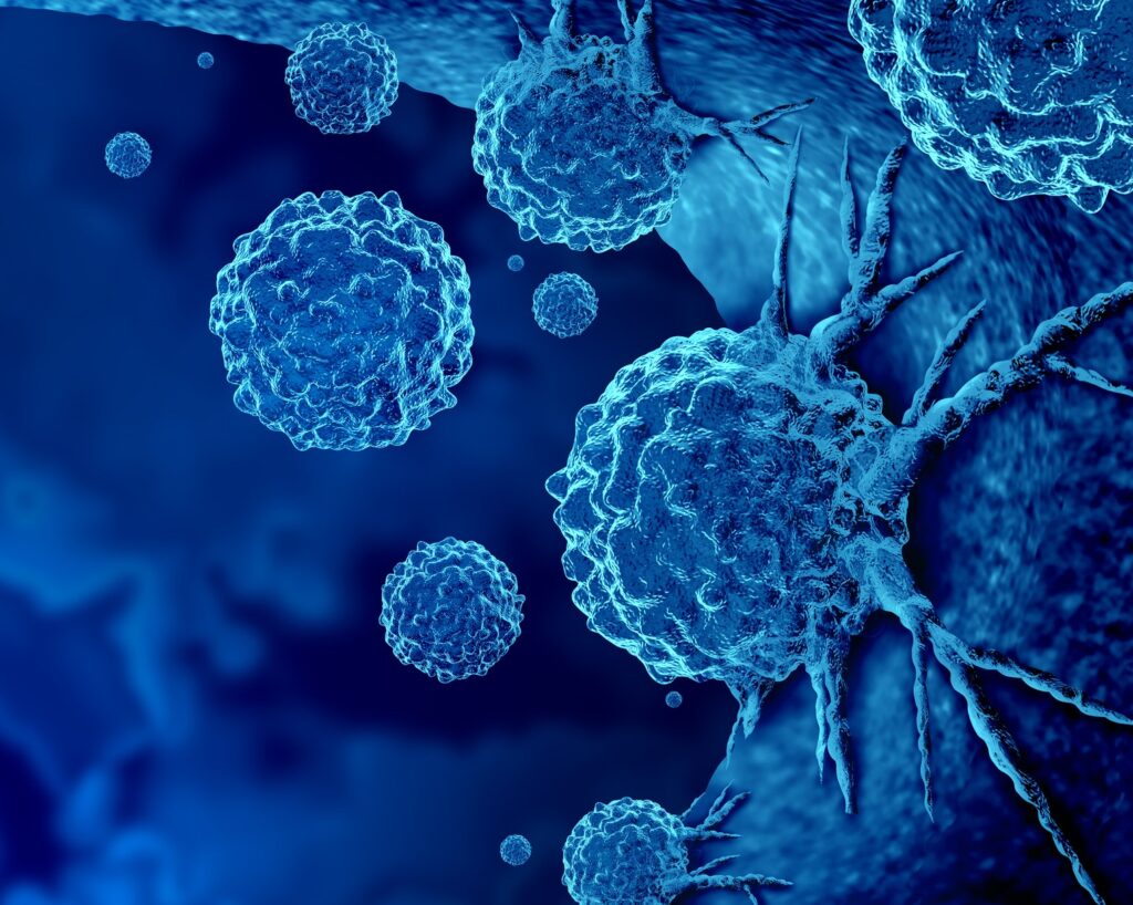
Despite decades of research, there remains a lack of effective and safe treatments for many forms of cancer, partially due to a lack of human-relevant models of cancer progression. Although animal models can effectively model some human cancer behaviors, they do not allow researchers to study the impact of specific tissue components, investigate the role of the tumor microenvironment (TME), or visualize how cancer cells behave over time when interacting with stromal and immune cells. Meanwhile, conventional in vitro models fail to reproduce the complexity that influences cancer behavior in vivo, and cannot appropriately model more recent advancements in cancer therapeutics.
How we can help
Model human-specific cancer therapies with Organ-on-a-Chip technology
Organ-on-a-Chip technology allows researchers to recreate the human tumor microenvironment in vitro, enabling mechanistic studies of cancer cell behavior and drug efficacy and safety. Endothelial co-culture provides important cell-cell interactions, while media flow and tissue-relevant stretch help model the mechanical forces cancer cells experience in the body. With these highly-tunable models, it is possible to modulate various cellular, molecular, chemical, and biophysical properties in a controlled manner to investigate their impact on cancer progression and behavior. The improved gene expression demonstrated in Organ-Chips also enables the evaluation of immuno-oncology therapeutic safety risk, not possible with conventional cell culture.
TUMOR MICROENVIRONMENT
Complex 3D in vitro models supporting novel interrogation of cancer progression
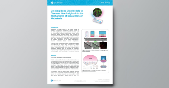
Cancer progression is a complex process that is not fully understood due to the limited ability of animal and in vitro models to recapitulate the human tumor microenvironment. With Organ-Chips, researchers can model early stages of cancer metastasis and intravasation of tumor cells into the vasculature. By modeling and tuning the tumor microenvironment (TME), including peristaltic-like stretch, media flow, and tumor-stromal tumor-endothelial interactions, researchers can better understand—and ultimately control—aspects of cancer progression.
Immuno-Oncology
Model the entire journey of CAR T-cell therapy for solid tumors, from vascular administration to target cell killing
With the CAR T workflow for Emulate Organ-Chips, researchers can model the entire journey of CAR T-cell therapy from blood vessel to target—including CAR T cell vascular administration, tumor inflammation-specific endothelial cell attachment and migration, and antigen-specific killing—in a vascularized cancer cell line model.
T-Cell Engagement and Activation
An emerging new standard for immunotherapeutic safety testing and optimization
Although promising for immunotherapy, T-cell bispecific antibodies (TCBs) present an on-target, off-tumor safety risk due to low levels of expression of tumor antigens in healthy tissues. Increasingly, TCBs target human-specific antigens not expressed in animal models, rendering them uninformative for preclinical studies. Even conventional human cell-based models fail to replicate in vivo gene expression, limiting their value. Because Organ-Chips incorporate human cells, cell-cell interactions, and mechanical forces, gene expression is much closer to in vivo, enabling a more human-relevant and clinically translatable assessment of TCB safety and efficacy.
User Applications
Cancer Case Studies
See how our user community is applying Organ-on-a-Chip technology to study everything from the tumor microenvironment to immuno-oncology.