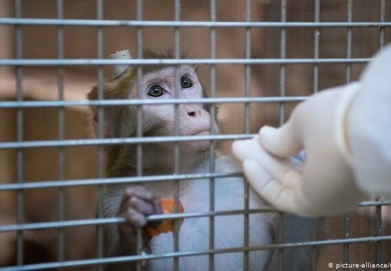
Earlier this week, The New York Times ran an article outlining the need to build a strategic monkey reserve to address shortages in rhesus monkeys that have arisen in the testing of COVID-19 vaccines. Shortages have arisen since the import of animals from China have been banned during the pandemic, causing prices of monkeys to skyrocket. But the article fails to address the real issue—it’s time to say, ‘enough is enough’ and begin to discourage the use of primates for drug testing. Our societal goal should be reducing the number of monkeys used and encouraging more innovative alternatives to primate testing, not breeding more laboratory animals to keep up with the high demand from drug developers. 

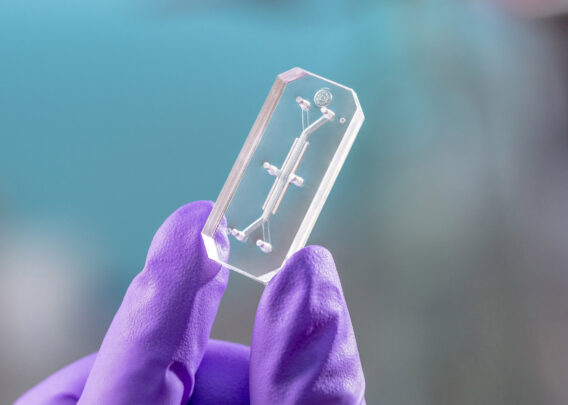
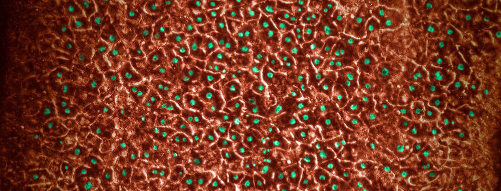
Beyond the obvious ethical concerns, animal testing has severe limitations in effectiveness, particularly for today’s novel biologic drugs and vaccines. Monkeys are not always capable of replicating human disease processes or drug response. Advances in biopharmaceutical research, including Organ-on-a-Chip technology, which recreates human biology in highly controlled lab settings, can more closely emulate human drug response. 

While animal testing may always play a role in some pharmaceutical testing, a strategic monkey reserve is not the answer. It’s our responsibility to explore alternatives. If better patient outcomes are the end game, we should encourage the use of technology that gets us there faster, and more humanely.   We are, however, starting to see progress from a policy perspective in recognizing the importance of new alternatives to primate (and other animals) testing. In 2020, Congressional Representatives Alcee L. Hastings (D-Florida) and Vern Buchanan (R-Florida) together introduced legislation that would help improve animal testing transparency, while incentivizing the study of more human-relevant testing methods. We hope that similar legislation is introduced into the new Congress, as we believe it is the first step towards replacing animal testing with new methods that can more effectively recreate human cellular responses.

Before a drug comes to market, it is subjected to rigorous tests to ensure its safety and efficacy. However, many standard safety tests for drug candidates involve rodent and non-rodent animal models which cannot always accurately predict which drugs might cause drug-induced liver injury (DILI) in humans. To increase predictive confidence ahead of clinical studies, a species-specific, multicellular, in vitro hepatotoxicity assay was developed, using our Liver-Chip, to characterize human toxicities, including responses that may have not been accurately predicted by animal models.

SNAPSHOT

Research Area: Hepatotoxicity testing

Organisms: Human, dog, rat

Sample Types: Human, dog, rat Hepatocytes, liver sinusoidal endothelial cells, Kupffer cells, and hepatic stellate cells

Research Question: Can a multispecies Liver-Chip predict liver toxicity and inform human relevance of liver toxicities detected in animal studies?

EXPERIMENTAL OVERVIEW

Emulate Organs-on-Chips technology was used to create rat, dog, and human Liver-Chips. Primary rat, dog, or human hepatocytes were seeded in the upper parenchymal channel within an extracellular matrix (ECM) layer on top of an ECM-coated, porous membrane that separates the two parallel microchannels. Species-specific rat, dog, or human liver sinusoidal endothelial cells (LSECs), with or without Kupffer cells and/or stellate cells, were cultured on the opposite side in the lower vascular channel. These quadruple-cell chips were characterized by albumin secretion and Cytochrome P450 enzymatic activity prior to drug toxicity testing.

A diagram of a Quad-Culture Liver-Chip

RESULTS

Studying species-specific drug toxicities
To explore whether the rat, dog or human liver chips could be used to predict species-specific DILI responses, the study evaluated hepatotoxic effects induced by bosentan, a dual endothelin receptor antagonist. Due to species differences in the bile salt export pump (BSEP) efflux transporter, bile salts accumulate within hepatocytes upon exposure to bosentan, causing cholestasis in humans, but not in rats or dogs. The results showed that the mechanisms of drug induced liver injury can be dissected using the Liver-Chip. 

Detection of diverse phenotypes of hepatotoxicity
The species-specific, quadruple cell Liver-Chips were tested with tool compounds that are known to cause different kinds of toxicity and provided insights into the drug mechanism of action. In brief, the experiments were designed to detect Kupffer cell depletion, and markers of steatosis and fibrosis, with the results suggesting that the Liver-Chip could be a useful model for predicting toxicities. In addition, candidate drugs that show hepatotoxicity in dog and rat models can be screened for potential risk assessment in the human quadruple cell Liver-Chip.

Towards a more accurate model for pre-clinical screening
One of the most difficult forms of hepatotoxicity to predict relates to DILI responses that are often missed during preclinical and early clinical testing. The human Liver-Chip was exposed to TAK-875, a G protein coupled receptor 40 agonist that was discontinued in phase 3 trials due to DILI in a few individuals. The experiments showed that continuous and prolonged exposure caused mitochondrial dysfunction, oxidative stress, formation of lipid droplets, and an innate immune response, all of which are harbingers of DILI for susceptible patients. These results highlight the advantage of the Liver-Chip for assessing the pathophysiological consequence of reactive metabolite formation, which has been strongly associated with DILI.

CONCLUSION

This study explores whether species-specific, in vitro hepatotoxicity assays using Liver-Chips can provide a greater understanding of the mechanisms of hepatotoxicity in animal models, which may lead to reduction in the number of animal studies to provide accurate risk assessment for drug candidates. The Liver-Chip detected diverse types of liver toxicity, including hepatocellular injury, steatosis, cholestasis, and fibrosis. The study has shown that species-specific Liver-Chips have potential future applications for hepatotoxicity testing, disease modeling, biomarker identification. This experimental approach could also be used to study whether toxicities observed in animal models are translatable to humans, and to understand human cellular variability with regards to drug response.  

REFERENCE

Jang, KJ et al. Reproducing human and cross-species drug toxicities using a Liver-Chip. Sci Transl Med 019 Nov 6;11(517).

A summary of the scientific paper published in Advanced Science, “Is it Time for Reviewer 3 to Request Human Organ Chip Experiments Instead of Animal Validation Studies?

Animal studies are an old practice, going back to the ancient Greeks, continuing with William Harvey and others in the 16th and 17th centuries and gaining greater traction in the past 100 years. Animal models offer more complexity than simple cell cultures, though they fall short of human physiology. But even with their known shortcomings, animal studies have been the gold standard for preclinical research – until now. 

To a large degree, the preference for animal studies was by necessity. Preclinical animal models were the best available research tools. However, in recent years, microfluidic Organ-Chips and other technologies have begun to offer superior alternatives. Because they use human tissues – in some cases cultured from patients – they can more accurately recapitulate many of the mechanisms associated with human physiology and bring us closer to personalized medicine.  

As many as 90% of all drugs that enter human clinical trials fail to gain FDA approval. Sometimes they are not effective and often, they are just too toxic. Our preclinical models should be able to catch these deficits before the drugs reach patients. However, the current system produces human suffering and wastes valuable time – we must identify better approaches. 

THE PROMISE OF ORGAN-CHIPS 

Gif of intestine peristalsis. It shows the biology expanding and contracting.

Microfluidic Organ-Chips offer a superior model because they can be built with human tissues and incorporate key physiological components, such as a vasculature, tissue-tissue interfaces, circulating immune cells, and even a complex microbiome. Mechanical forces, such as those associated with breathing or peristalsis, can be replicated as well. Advanced fluidics bring in nutrients, remove waste, and permit mimicry of dynamic changes in drug levels over time, better recapitulating what patients experience in vivo

These chips are transparent and small, which means researchers can observe reactions continuously, in real time, through microscopes or more advanced systems – something that cannot be done in animal models.  

But most importantly, from a drug development standpoint, these chips have been proven to provide important windows into human physiology and provide greater insight into how specific cells and tissues respond to therapeutic molecules. For example, lung chips have replicated human COPD and non-small cell lung cancer. Organ-chips have also modeled intestines, blood-brain barrier, bone marrow, liver, kidneys and numerous other organs and tissues. 

Organ-Chips can be particularly useful when investigating human immune responses. Different species of mammals have different immune systems, which can make them ineffective models when studying vaccines, cancer immunotherapies or autoimmune disease treatments.

One chip, modeling a single organ system, can be quite powerful. But these devices also can be linked fluidically for multi-organ studies, creating even more sophisticated “body-on-chips” models. These systems can incorporate many of the complex inter-organ signaling mechanisms found in the human body – for example, pancreatic islet cells can talk to liver tissue. Investigators can even model many of the complexities associated with the reproductive system.

THE COST OF FAILURE

The computer industry has a saying – garbage in, garbage out – and that could easily be applied to the biopharmaceuticals drug development pipeline. Inadequate preclinical models translate into failed drugs. If this were a purely academic discussion, the industry could be forgiven for moving slowly. But these poor models have real world ramifications when ineffective and/or toxic drugs enter human trials where at best they fail to deliver hoped-for benefits or worse, cause patients physical harm.

Organ-Chips have proven themselves over and over, identifying toxicities that went previously undetected in animal studies but unfortunately showed up in human trials. In one instance, a Blood Vessel-Chip retrospectively revealed the faults in a monoclonal antibody therapy that led to blood clot formation in the lung. Unfortunately, the drug had already caused deaths in the human study.  

Better preclinical reads on efficacy and safety mean drugs that would have been likely to fail in human trials can be shelved or redesigned. Medicines that work best in subpopulations can be tested early in those populations, rather than failing in a large trial and being reapplied to the smaller group as a consolation prize. Ultimately, making the system more efficient could accelerate our ability to conduct successful clinical trials, dramatically reducing costs and bringing new therapies to patients faster.

But it’s not just about drugs that show promise in animal models and fail in humans. We must also assume there are molecules that fail in animals but are safe and effective in people. However, we approach this problem, it’s obvious we are leaving good drugs on the table.

This is not to say that Organ-Chips are perfect models. They have trouble replicating peripheral neurons, fat and other tissues, which could generate serious blind spots. More work needs to be done to help these emerging research tools better incorporate in vivo human physiology. And that’s really the point. We can no longer be satisfied with the flawed preclinical models that have produced such poor results. Under the current regime, unmet medical needs, such as Alzheimer’s disease, continue to be unmet, and patients, caregivers and communities suffer. Microfluidic Organ-Chips will not solve all these issues, but they will provide insights animal models do not and will ultimately bring a more rational approach to our preclinical discovery efforts.