From July 3rd–5th, 2024, Emulate joined the European Organ-on-Chip society (EUROoCS) annual conference hosted at the historical Leonardo Campus of Politecnico di Milano in the splendid city of Milan, Italy.
What is EUROoCS?
EUROoCS is an independent, not-for-profit organization established to encourage and develop research into Organ-Chips and provide opportunities to share knowledge and advance the field with the ultimate goal of creating a healthier future. EUROoCS holds an annual conference fostering worldwide delegates active in the field of Organ-on-a-Chip technology and microphysiological systems to showcase the latest scientific breakthroughs and engage in meaningful discussions on the field’s cutting-edge advancements. The conference brings together a broad audience of Organ-Chip experts and enthusiasts from government, academia, pharma, medicine, and other institutions.
Learn how you can join EUROoCS here.
EUROoCS in Review
This exciting event started with the plenary talk from Prof Andries D. van der Meer, EUROoCS Chair, highlighting the “Official publication of the international Roadmap for Organ-on-Chip Standardization.” This was followed by a keynote talk titled, “Building a roadmap towards Regulatory Acceptance of NAMs in the Development and Approval of Pharmaceuticals,” delivered by Sonja Beken, PhD, Coordinator of the Unit of non-clinical evaluators at the Belgian Federal Agency for Medicines and Health Products (FAMHP), which highlighted the vision of the 3Rs Working Party and Member of the Non-Clinical Working Party at the European Medicines Agency. There was also a consecutive regulatory industrial round table discussion on Organ-Chip-based test methods for the authorization of medicinal products. Another interesting keynote was delivered by Kimberly Homan, PhD, where she discussed how combining organoids with Organ-Chips opens new doors in drug research and development.
Another interesting keynote was delivered by Genentech’s Kimberly Homan, PhD, where she discussed how combining organoids with Organ-Chips allows researchers to unlock new possibilities in drug research and development.
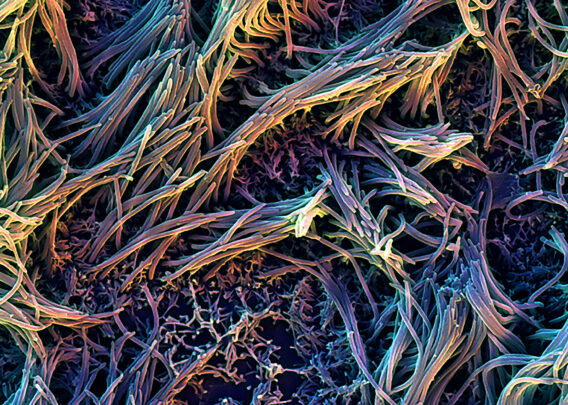
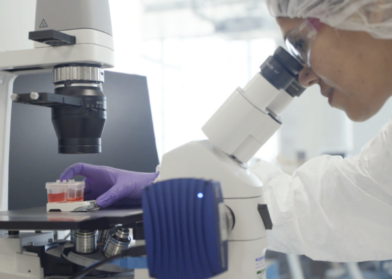
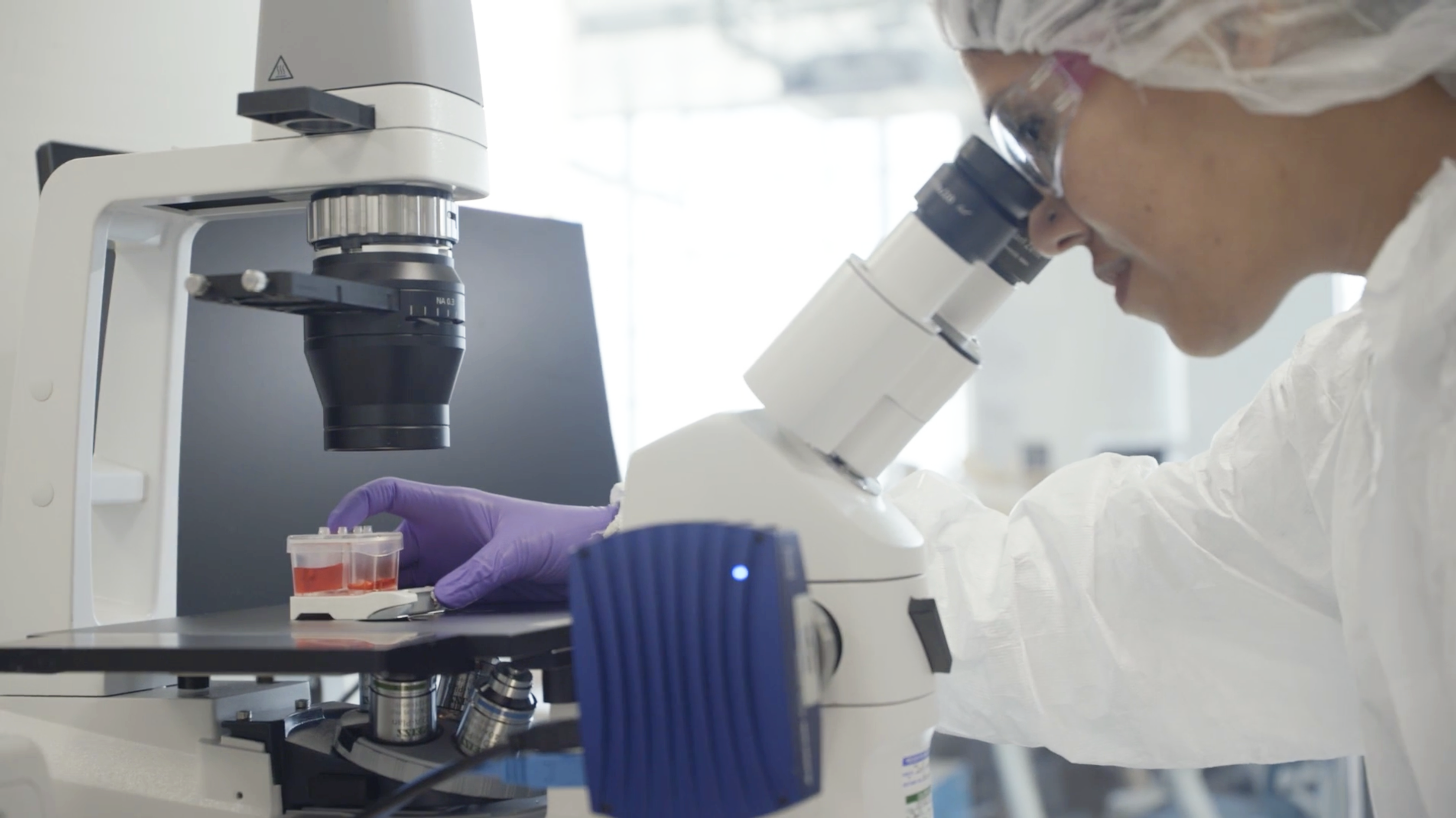
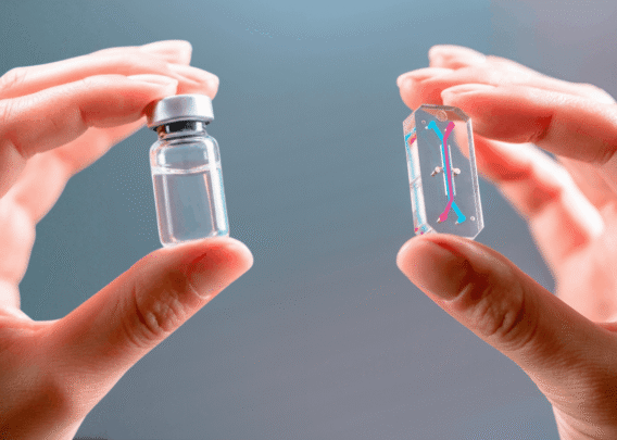
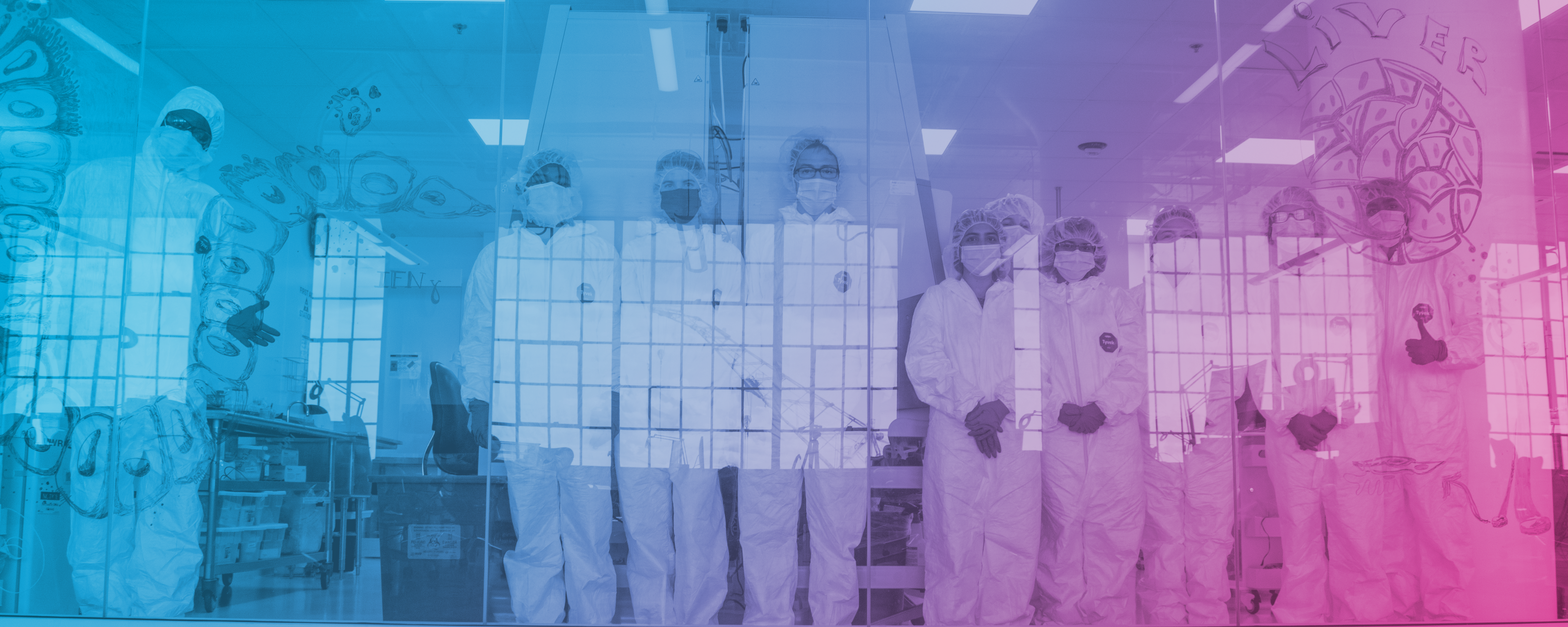
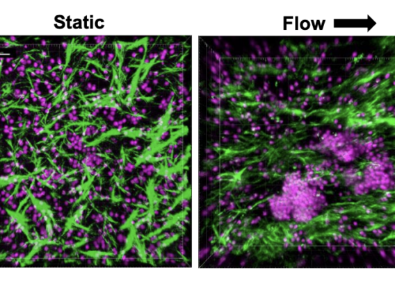
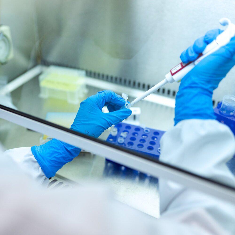
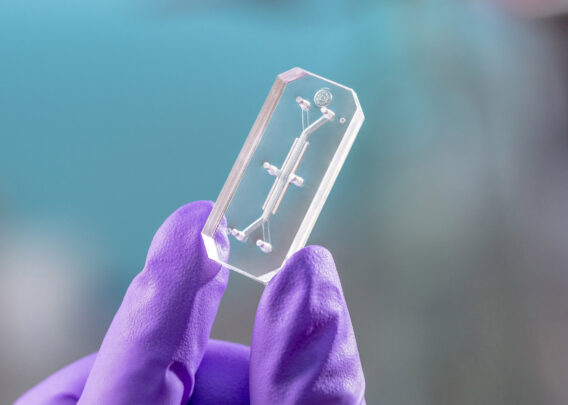
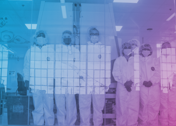
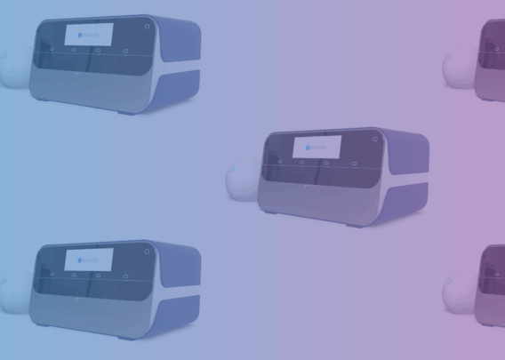
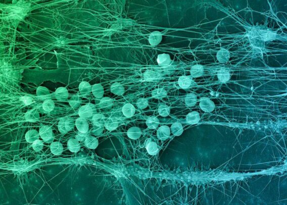
At the conference, Emulate provided attendees an opportunity to gain hands-on experience in our Organ-Chip wet lab and explore cutting-edge innovations, including the Chip-A1 Accessible Chip and our new CAR T Organ-Chip application.
Our customers also showcased their latest scientific breakthroughs in presentations and poster sessions.
Talks:
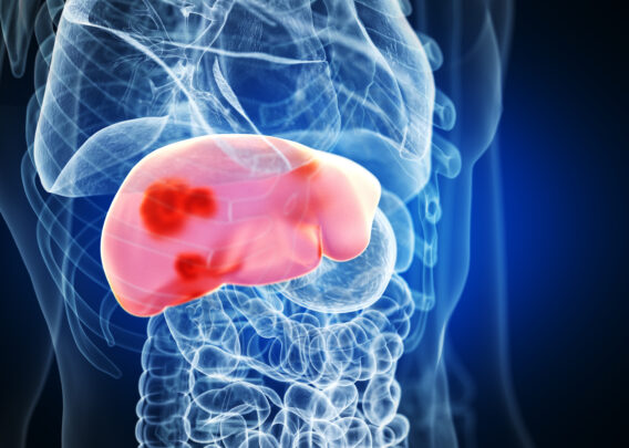
- “Comparison of Spheroids and a Microfluidic Chip Model as Advanced in Vitro Liver Models” by Heidrun Ellinger-Ziegelbauer, PhD, Principal Scientist, Bayer AG Pharmaceuticals
- “Recapitulating memory B cell responses in a Lymphoid Organ-Chip to evaluate mRNA vaccine boosting strategies” by Lisa Chakrabarti, PhD, Control of Chronic Viral Infections Group, Virus and Immunity Unit, Institute Pasteur, Université de Paris
- “Development and validation of an organ-chip model of breast cancer bone metastasis” by Stefaan Verbruggen PhD, Centre for Predictive in vitro Models, Engineering and Materials Science, Queen Mary University of London, London, United Kingdom
Posters:
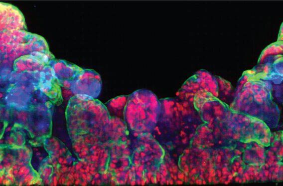
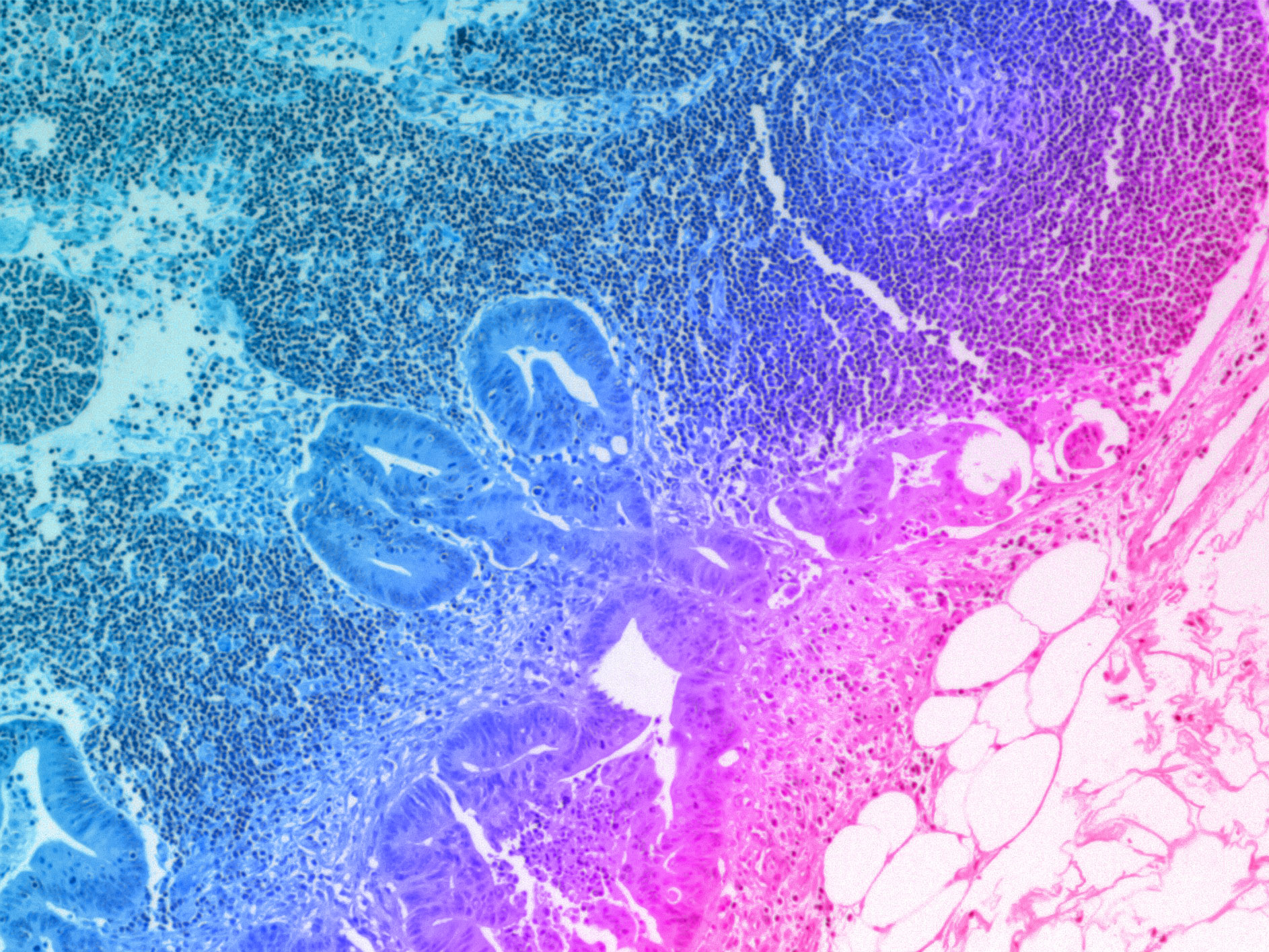
- “Impact of tissue microenvironment and mechanical forces of the human gut on pathogens invasion” by Samy Gobaa, PhD, Head of Organ-on-Chip Center, Institute Pasteur
- “Lung organoid-on-chip models to study respiratory infections” by Barbara Fonseca, PhD, Researcher, Institut Pasteur
- “Evaluation of Cell-Type Specific miRNA Secretion in a Liver-Chip Model” by Nicole Reisinger, DSM-Firmenich, ANH R&D Center, Tulln, Austria
EUROoCS2024 was an amazing opportunity to connect with fellow Organ-Chip enthusiasts and see how scientists are using Emulate Organ-on-a-Chip technology to advance research across a wide variety of areas. The field of Organ-Chips is in a better place than it’s ever been, and we’re excited to see what new achievements will be made over the next year. See you at EUROoCS2025!
__________________
Explore past and future Emulate events here.
Discover the exciting work of our user community by downloading our Publication Digest.