Webinar Abstract
Featured session at Boston MPS Day, which took place on 11/1/2023.
Chimeric antigen receptor (CAR) T-cell therapy holds great promise as a treatment for a variety of human cancers. While there are 5 FDA-approved CAR T therapies for liquid tumors, successfully adapting this treatment for solid tumors remains elusive.
A major reason why solid tumors are so much harder to treat with CAR Ts is because of their vastly different microenvironment. To effectively kill solid tumor cells, CAR Ts must first exit the vasculature at the tumor location, infiltrate deeply into the tumor, lock in with tumor-specific antigens, and finally, kill the cancer cells.
It’s a long, complicated journey, and one that conventional models cannot capture due to their simplicity. Enter Organ-Chips.
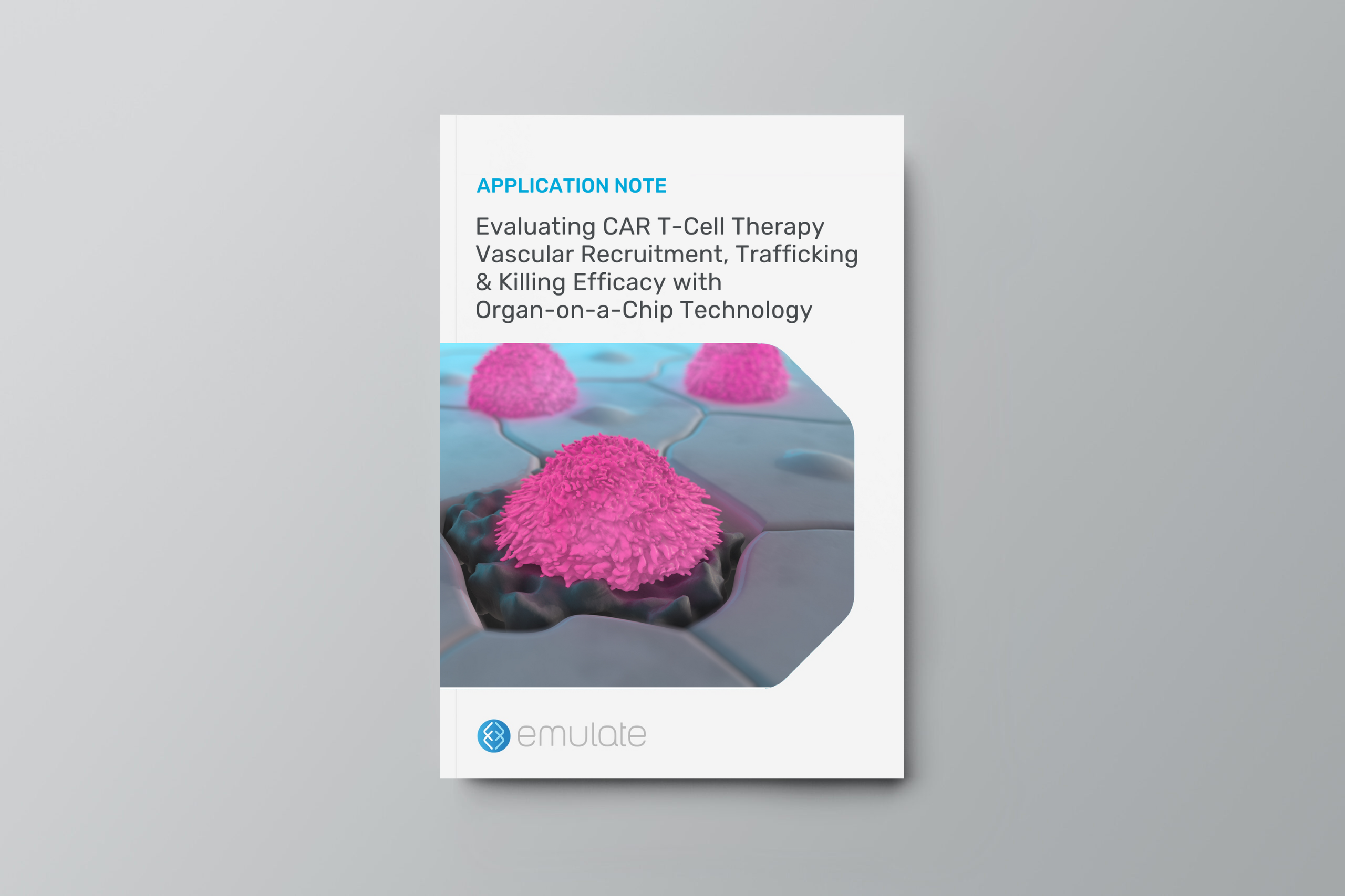
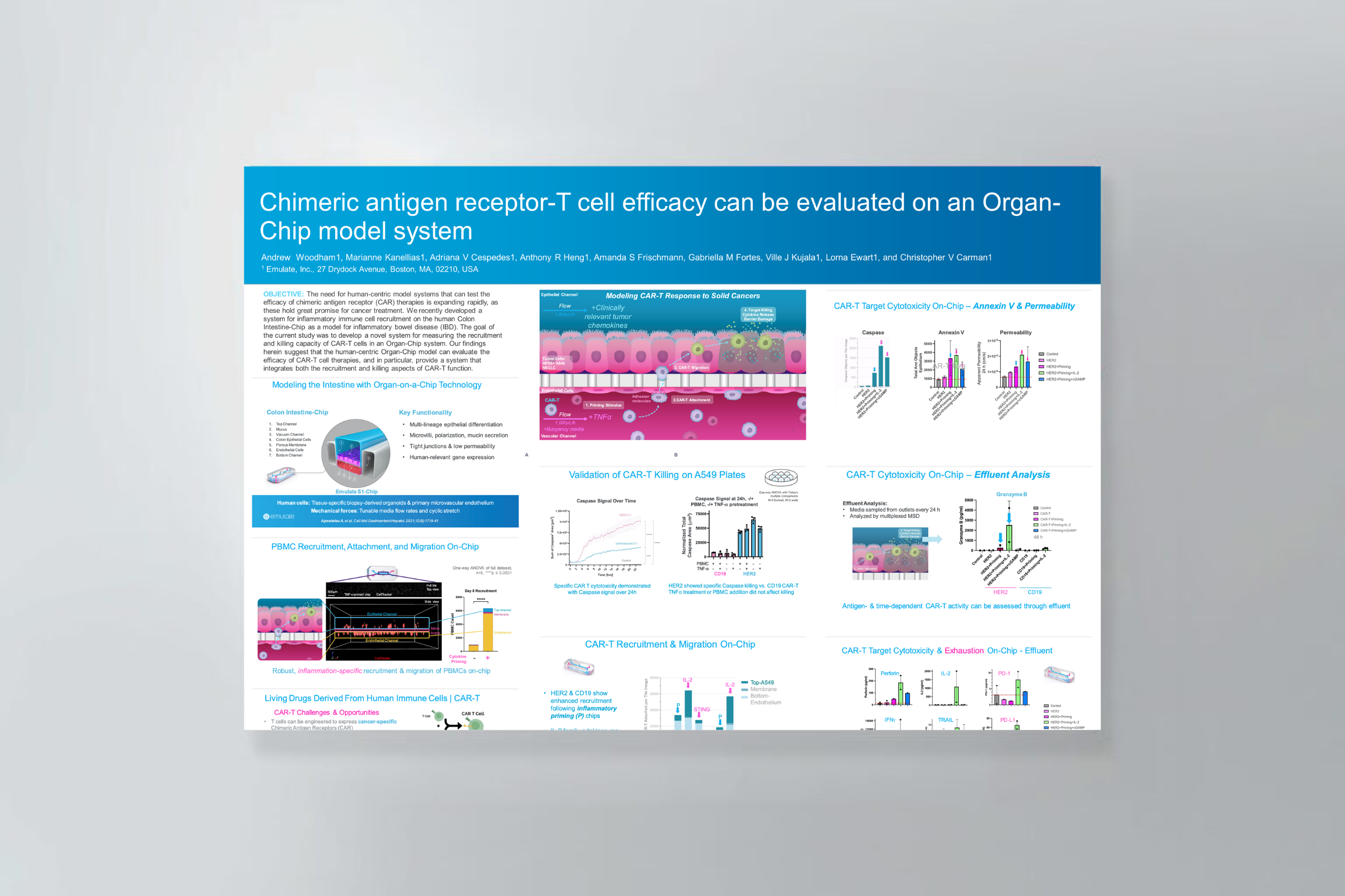
Emulate Organ-Chips can help address these challenges by enabling human-relevant and more comprehensive recapitulation of immunotherapy efficacy. In this webinar, Chris Carman, PhD, shared data from proof-of-concept studies showing how Organ-Chips can be used to evaluate the efficacy of CAR T-cell therapies and provide a system that integrates both the recruitment and killing aspects of CAR T function.
In this webinar, you will learn how Organ-Chips can be used to:
Oncology drug candidates are currently the least likely type of therapeutic to succeed in clinical trials, with only 5.1% of Phase I candidates going on to receive FDA approval1. Understanding a tumor’s microenvironment is key to regulating cancer progression and developing more effective therapies—and Chip-A1 will give researchers this capability.
In this August 15, 2023 webinar, Luke Dimasi, Senior Director of Product Management at Emulate, provided an overview of the Chip-A1 Accessible Chip, a new Organ-Chip consumable that expands the applications of Organ-on-a-Chip technology by allowing users to create thicker, multilayered tissues within the epithelial culture chamber and directly treat the tissue with topical or aerosolized drugs. Following this introduction, Elee Shimshoni, PhD, postdoctoral researcher at MIT, discussed how she and her former team from the Wyss Institute at Harvard used a prototype of Chip-A1 and found that it offered a new approach for studying epithelial-stromal interactions in Barrett’s Esophagus as well as the broader underlying mechanisms associated with esophageal cancer progression.
During this webinar, the speakers discussed:
About Organ-on-a-Chip Technology
Organ-on-a-Chip technology is poised to deliver a paradigm shift in drug discovery. By emulating human physiology, Organ-Chips have the potential to increase the predictive power of preclinical modeling and advance more drugs to the clinic. Learn more about Organ-on-a-Chip technology by downloading our free eBook.
The need for human-centric model systems that can test the efficacy of chimeric antigen receptor (CAR) therapies is expanding rapidly, as these hold great promise for cancer treatment. We recently developed a system for inflammatory immune cell recruitment on the human Colon Intestine-Chip as a model for inflammatory bowel disease (IBD). The goal of the current study was to develop a novel system for measuring the recruitment and killing capacity of CAR-T cells in an Organ-Chip system.
Our proof-of-concept findings herein suggest that the human-centric Organ-Chip model can evaluate the efficacy of CAR-T cell therapies, and in particular, provide a system that integrates both the recruitment and killing aspects of CAR-T function.
Over the past 15 years, organoids have revolutionized the study of human organ and tumor behavior. In the coming years, Organ-on-a-Chip technology promises to rapidly increase the complexity of human organotypic models, enabling the discovery of more physiologically relevant insights into human health and disease.
In this on-demand webinar, Jens Puschhof, PhD, of the German Cancer Research Center (DKFZ) discusses how lessons learned from organoid biology can be applied to Organ-on-a-Chip research and examine areas where these two technologies are being combined with great potential for synergy. In particular, he shares aspects of his team’s work studying the impact of cancer-associated bacteria in colorectal cancer metastasis using Emulate Organ-Chip models.
Key discussion points / learnings:
Organ Model: Esophagus
Application: Cancer
Abstract: The pathogenesis of subsquamous intestinal metaplasia (SSIM), in which glands of Barrett’s esophagus (BE) are buried under esophageal squamous epithelium, is unknown. In a rat model of reflux esophagitis, we found that columnar-lined esophagus developed via a wound-healing process involving epithelial-mesenchymal plasticity (EMP) that buried glands under ulcerated squamous epithelium. To explore a role for reflux-induced EMP in BE, we established and characterized human Barrett’s organoids and sought evidence of EMP after treatment with acidic bile salts (AB). We optimized media to grow human BE organoids from immortalized human Barrett’s cells and from BE biopsies from seven patients, and we characterized histological, morphological, and molecular features of organoid development. Features and markers of EMP were explored following organoid exposure to AB, with and without a collagen I (COL1) matrix to simulate a wound-healing environment. All media successfully initiated organoid growth, but advanced DMEM/F12 (aDMEM) was best at sustaining organoid viability. Using aDMEM, organoids comprising nongoblet and goblet columnar cells that expressed gastric and intestinal cell markers were generated from BE biopsies of all seven patients. After AB treatment, early-stage Barrett’s organoids exhibited EMP with loss of membranous E-cadherin and increased protrusive cell migration, events significantly enhanced by COL1. Using human BE biopsies, we have established Barrett’s organoids that recapitulate key histological and molecular features of BE to serve as high-fidelity BE models. Our findings suggest that reflux can induce EMP in human BE, potentially enabling Barrett’s cells to migrate under adjacent squamous epithelium to form SSIM.NEW & NOTEWORTHY Using Barrett’s esophagus (BE) biopsies, we established organoids recapitulating key BE features. During early stages of organoid development, a GERD-like wound environment-induced features of epithelial-mesenchymal plasticity (EMP) in Barrett’s progenitor cells, suggesting that reflux-induced EMP can enable Barrett’s cells to migrate underneath squamous epithelium to form subsquamous intestinal metaplasia, a condition that may underlie Barrett’s cancers that escape detection by endoscopic surveillance, and recurrences of Barrett’s metaplasia following endoscopic eradication therapy.
To learn more about these findings, view our webinar “Organ-Chips 201: The Importance of Flow, Stretch, and Stroma for in vitro Modeling.”
Originally presented at Global MPS Day 2023
In this on-demand webinar, Samaneh Kamali, Ph.D., of Champions Oncology Learn how Champions Oncology is accelerating oncology drug development by building complex in vitro models with patient-derived TumorGrafts.
Article Type: Review
Application: Cancer
Abstract: Organ-on-chip systems are capable of replicating complex tissue structures and physiological phenomena. The fine control of biochemical and biomechanical cues within these microphysiological systems provides opportunities for cancer researchers to build complex models of the tumour microenvironment. Interest in applying organ chips to investigate mechanisms such as metastatsis and to test therapeutics has grown rapidly, and this review draws together the published research using these microfluidic platforms to study cancer. We focus on both in-house systems and commercial platforms being used in the UK for fundamental discovery science and therapeutics testing. We cover the wide variety of cancers being investigated, ranging from common carcinomas to rare sarcomas, as well as secondary cancers. We also cover the broad sweep of different matrix microenvironments, physiological mechanical stimuli and immunological effects being replicated in these models. We examine microfluidic models specifically, rather than organoids or complex tissue or cell co-cultures, which have been reviewed elsewhere. However, there is increasing interest in incorporating organoids, spheroids and other tissue cultures into microfluidic organ chips and this overlap is included. Our review includes a commentary on cancer organ-chip models being developed and used in the UK, including work conducted by members of the UK Organ-on-a-Chip Technologies Network. We conclude with a reflection on the likely future of this rapidly expanding field of oncological research.
Colorectal cancer (CRC) is one of the deadliest cancers worldwide with over 900,000 people dying from the disease each year (Siegel et al., 2021). In the United States, the 5-year survival rate for patients with metastatic CRC is less than 15% (Siegel et al., 2020). To address this dismal outcome, there is an urgent need to better understand and ultimately control aspects of cancer progression.
In a recent paper published in iScience, researchers from the Lawrence J Ellison Institute for Transformative Medicine describe an in vitro Organ-Chip model that emulates in vivo tissue structure and the tumor microenvironment (TME) to better understand intravasation, an early step in metastasis.
In this on-demand webinar, Dr. Mumenthaler discusses recent advancements made through combining Organ-Chip models with high content imaging and mass spectrometry-based metabolomics to improve our understanding of microenvironmental contributions to colorectal cancer progression.
Study results that will be discussed:
Organ Model: Intestine (Colorectal cancer)
Application: Cancer
Abstract: Despite colorectal cancer’s (CRC) prevalence, its progression is not well understood. The microfluidic organ-on-chip (OOC) model described herein recreates the epithelial-endothelial tissue-tissue interface, fluid flow, and mechanical forces that exist in vivo, making it an attractive model to understand and ultimately disrupt CRC intravasation. This protocol provides step-by-step details for tumor cell seeding to create a CRC-on-chip model, chip effluent collection and analysis, and on-chip imaging to monitor tumor cell invasion within a more physiologically relevant microenvironment. For complete details on the use and execution of this protocol, please refer to Strelez et al. (2021).