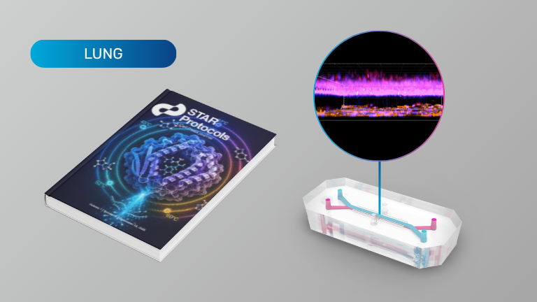
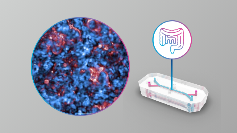
Protocol for generation of a 3D humanized lung-tumor-on-a-chip model of immune-hot and immune-cold microenvironments

Organ Model: Lung

Application: Cancer

Researchers created a fully humanized lung tumor microenvironment that models both immune-hot and immune-cold tumors. Human lung cancer cells were cultured in one channel while primary lung microvascular endothelial cells formed a perfused vascular compartment in the other, enabling researchers to recreate key tumor–vascular interactions under flow. Human peripheral blood mononuclear cells were then introduced through the vascular channel to study immune cell recruitment and migration toward the tumor, with responses characterized using immunofluorescence imaging, flow cytometry, and secretome analysis. The protocol provides a platform for investigating how tumor, endothelial, and immune cells interact within the tumor microenvironment and can potentially be adapted to other cancer types.

Products Used In This Publication

Emulate Community Publications Digest: Summer 2026 Issue

With over 150 peer-reviewed publications across 35+ organ models, Emulate Organ-Chips are empowering researchers to make game-changing scientific breakthroughs! Download this digest to easily explore all publications related to your field of research, or to just learn more about how the technology itself is developing.

New this quarter:

Fallopian Tube

  • Human fallopian tube-on-a-chip for preclinical testing of non-hormonal contraceptives with living human sperm

Intestine

  • Probiotic intervention mitigates radiation-induced intestinal injury by alleviating oxidative stress in a human gut-on-a-chip

Lung (Airway)

  • Development of an acute inhalation toxicity testing method based on a lung-on-a-chip

Lung (Alveolus)

  • Enhanced lung delivery of an immunostimulatory duplex RNA augments the antitumor activity by reshaping systemic cytokine pharmacodynamics

Lung (Alveolus)

  • Alveolus-on-a-Chip: A Novel Tool for Modeling Lung Transplant Cold Storage Ischemia/Reperfusion Injury

Placenta

  • Simvastatin Restores Uteroplacental Hemodynamics and Trophoblast Function in Obstetric Antiphospholipid Syndrome in a Placenta-on-a-Chip Model

REVIEW ARTICLES:

Female Reproductive Tract

  • Female Reproductive Tract Organ-on-Chips: Modeling Barrier Function and Drug Transport

Drug Development

  • Human organ-on-a-chip technology as a catalyst for drug discovery

PUBLISHED FROM PRE-PRINT:

Intestine (Colon)

  • Human inflammatory bowel disease-on-a-chip for modelling disease progression, cancer initiation and sex-specific effects

Lymph Node

  • In vitro recapitulation of intramuscular mRNA vaccination with naive and recall antigens using a human lymphoid follicle chip platform

Vasculature

  • Human coronary artery organ-chip with circulating immune cells recapitulates anti-inflammatory effect of pulsatile wall strain

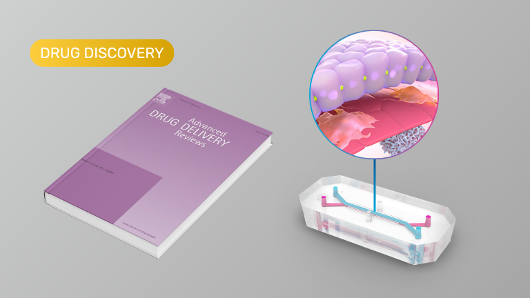
Human organ-on-a-chip technology as a catalyst for drug discovery (Review)

Application: Drug Discovery

This review highlights Organ-Chips as a more human-relevant preclinical platform that recreates key aspects of organ physiology—including tissue-tissue interfaces, dynamic fluid flow, mechanical forces, and immune interactions—to improve prediction of human drug responses. Unlike conventional cell cultures or animal models, Organ-Chips enable therapeutics to be evaluated using clinically relevant dosing routes and pharmacokinetic profiles, supporting more accurate assessment of efficacy, toxicity, and mechanisms of resistance. The authors also emphasize that combining Organ-Chips with high-throughput screening, functional genomics, and artificial intelligence could accelerate target identification and drug discovery while helping reduce the high rate of clinical trial failures. Although challenges remain around standardization and scalability, the review concludes that Organ-Chips are poised to become an increasingly important tool for drug development and regulatory safety assessment.

Enabling Drug Development with NAMs: Scalable Imaging and AI Analysis Workflows for Organ-Chips

Synopsis

Organ-on-a-Chip technology is emerging as a powerful New Approach Methodology (NAM) for drug development, driven by the need for more human-relevant and scalable experimental models. As these systems move toward broader adoption, a key challenge remains: generating consistent, interpretable data that supports confident experimental and translational decision-making.

This webinar examines how imaging and AI-driven analysis workflows enable Organ-Chip studies to scale from innovation to routine application. Speakers begin with an overview of Organ-on-a-Chip technology and its role in addressing translational gaps in drug discovery, highlighting how Liver-Chips are being evaluated in collaboration with regulatory agencies for better prediction of drug-induced liver injury.

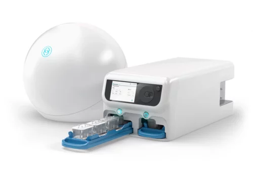
The session then explores how the newly released AVA™ Emulation System enables scalable Organ-Chip experimentation through an integrated system for incubation, microfluidic delivery, and routine imaging. Paired with AI-driven analysis, brightfield image data can be used to automate quality control by monitoring chip health, morphology, and assay performance over time across large studies.

To complete the workflow, post-study high-resolution imaging is applied to evaluate more complex biological markers, including toxicology-relevant endpoints and drug uptake. These datasets are paired with advanced analysis techniques that translate imaging data into quantitative, biologically meaningful insights.

Attendees will gain a practical understanding of how unified imaging and analysis strategies—spanning routine QC through advanced interrogation—support scalability, reproducibility, and alignment with evolving regulatory expectations for Organ-Chips and other NAM-based drug development.

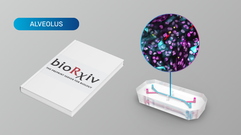
Enhanced lung delivery of an immunostimulatory duplex RNA augments the antitumor activity by reshaping systemic cytokine pharmacodynamics

Organ Model: Lung Cancer (Alveolus)

Application: Cancer, Immunotherapy

In this study, researchers used a human Lung Cancer-Chip to evaluate the therapeutic activity of a lung-targeted lipid nanoparticle (LungLNP) delivering an immunostimulatory duplex RNA (RNA-1) in a physiologically relevant human lung tumor microenvironment. The Lung Cancer-Chip recreated the alveolar interface by co-culturing human alveolar epithelial cells, pulmonary microvascular endothelial cells, and GFP-labeled A549 lung cancer cells under vascular perfusion, enabling intravenous-like dosing through the endothelial channel. Treatment with LungLNP/RNA-1 produced dose-dependent inhibition of tumor growth, stimulated secretion of interferons and pro-inflammatory cytokines, and revealed preferential uptake of the therapeutic by endothelial cells. These findings demonstrate the utility of Organ-Chips for evaluating targeted RNA therapeutics, elucidating mechanisms of cellular uptake and innate immune activation, and validating anti-tumor efficacy in a human-relevant tissue model.

Products Used In This Publication

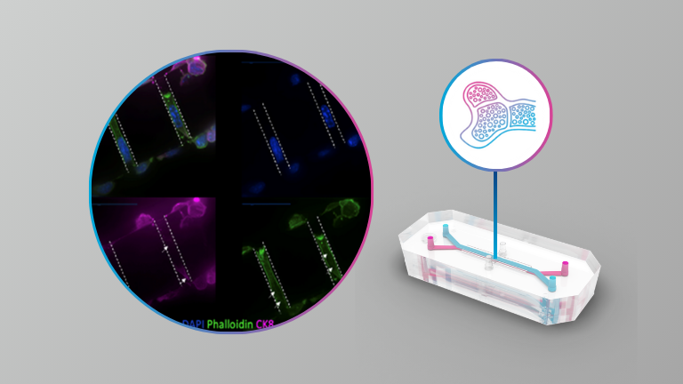
Multi-omics qualification of an organ-on-a-chip model of osteolytic bone metastasis

Organ Model: Bone (Breast cancer metastasis)

Applications: Cancer

Researchers recreated the osteolytic bone metastatic niche by co-culturing osteocytes, osteoclasts, and breast cancer cells in an Organ-Chip system. Multi-omics analyses—including RNA sequencing, cytokine profiling, and imaging—showed that the full tri-culture recapitulated in vivo biology, revealed synergistic pro-metastatic signaling, and aligned closely with gene expression profiles from gold-standard mouse models. These results validate this Organ-Chip model of osteolytic bone mestastasis as a scalable, human-relevant alternative to animal models and highlight its potential as a medium-throughput platform for uncovering therapeutic targets and screening drug candidates.

Products Used In This Publication

A New Paradigm for Personalized Cancer Medicine: Predicting Patient-Specific Therapeutic Responses with Organ-on-a-Chip Technology

Synopsis

Personalized medicine has long held the promise of tailoring treatments to the biology of each individual patient. Until now, however, the field has lacked a truly predictive human model. In a breakthrough that could transform how precision oncology is practiced, Dr. Lorenzo Ferri (McGill University) and Dr. Donald Ingber (Wyss Institute at Harvard University) have developed a patient-derived Organ-Chip model that accurately predicts individual responses to cancer therapeutics, outperforming current organoid models.  

In this webinar, Dr. Ferri, a leading clinician-scientist in esophageal and foregut malignancies, will share how his team leveraged Organ-on-a-Chip technology to create personalized models of EAC directly from patient tumors. These EAC-Chips faithfully recapitulate each patient’s unique tumor microenvironment and response to chemotherapeutics, marking a major advance toward functional precision oncology. 

Following the presentation, Dr. Ingber, the pioneer of Organ-on-a-Chip technology, will join Dr. Ferri for a live discussion and Q&A exploring how this work establishes Organ-on-a-Chip technology as a transformative platform for personalized medicine. Together, they will examine the implications of this study for clinical translation, therapeutic development, and the future of individualized cancer care. 

Key Learning Points 

  • Discover a new approach to personalized medicine: Learn how patient-derived Organ-Chips can replicate individual tumor biology and predict patient-specific therapeutic responses. 
  • Compare Organ-Chips and organoids: Understand why Organ-Chip models more accurately capture human tissue physiology and drug sensitivity than traditional organoid systems. 
  • Explore translational potential: See how Organ-on-a-Chip technology bridges clinical and experimental research, creating opportunities for functional precision oncology and more effective treatment selection. 
  • Gain insight from leaders in the field: Hear directly from the study’s senior authors on how integrating clinical expertise and bioengineering innovation can accelerate personalized cancer care. 

GABAergic signaling contributes to tumor cell invasion and poor overall survival in colorectal cancer

Organ Model: Colorectal cancer

Application: Cancer

This study used Organ-Chips to recreate patient-specific colorectal cancer tumors in a dynamic, gut-like environment. The chips revealed that cancers with Ras mutations produce excess GABA, which drives tumor invasion, and that blocking this pathway can reduce invasiveness. These insights link clinical trial data to a new therapeutic opportunity and highlight the power of Organ-Chips to uncover cancer mechanisms that traditional models miss.

A Patient-Derived Organ-on-Chip Platform to Model the Colorectal Tumor Microenvironment and Cancer Progression

In this webinar from Drug Discovery Day 2024, Carly Strelez, PhD, discusses her work using Organ-on-a-Chip technology to better understand colorectal cancer progression and transform approaches to personalized medicine.

Cancer-on-a-chip: Modeling Colorectal Cancer Progression

In this on-demand webinar, Shannon Mumenthaler, PhD, discusses recent advancements made through combining Organ-Chip models with high content imaging and mass spectrometry-based metabolomics to improve our understanding of ​microenvironmental contributions to colorectal cancer progression. 

Products Used In This Publication

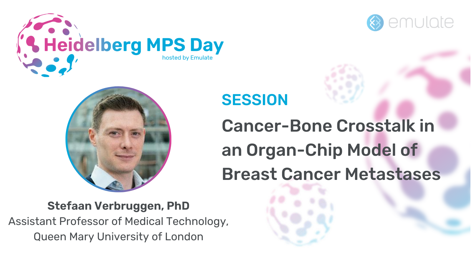
Cancer-Bone Crosstalk in an Organ-Chip Model of Breast Cancer Metastases

In this session from Heidelberg MPS Day, Dr. Stefaan Verbruggen presents his work on developing advanced Organ-Chip models to explore how breast and prostate cancer cells interact with bone tissue, particularly osteocytes. His research aims to understand why these cancers metastasize to bone and how the bone’s internal cells contribute to tumor growth. By combining cancer cells with osteoblasts, osteoclasts, and osteocytes in microfluidic chips and applying controlled fluid flow, his team can recreate key aspects of the bone environment. This approach reveals differences from traditional 2D cell culture methods, showing more complex, physiologically relevant behaviors. Ultimately, Dr. Verbruggen’s work seeks to refine these models, incorporate patient-derived cells, and achieve more accurate predictions of cancer progression and potential therapeutic responses.

Cancer and the Microbiome

This webinar focuses on the complex relationship between cancer development and the human microbiome, and how understanding this interplay might improve prevention and treatment strategies.

In the first half of the webinar, Dr. Rafik Fellague-Chebra, an executive global group medical director at Novartis with over two decades of experience in oncology, begins by reviewing the fundamental hallmarks of cancer—such as sustained proliferation, evading immune surveillance, and promoting angiogenesis. He introduces the concept of the “oncobiome,” describing how imbalances in gut microbes and other microorganisms can contribute to cancer initiation, progression, and response to therapy. He discusses how certain bacterial species and their metabolites may foster tumor growth and metastasis, while others could enhance immune responses or even improve the effectiveness of cancer treatments. Finally, he explores emerging therapeutic approaches—such as probiotics, fecal microbiota transplants, and personalized microbiome interventions—that may one day help predict, prevent, and more effectively treat cancer.

In the second half of the webinar, Dr. Jens Puschhof from the German Cancer Research Center focuses on leveraging advanced Organ-Chip models to dissect the impact of the microbiome on cancer progression. These engineered microfluidic platforms replicate key aspects of human organs and tumors, allowing precise control of the cellular environment and microbial composition. By integrating organoids derived from patient samples with specific microbial communities, his research illuminates the intricate cellular and molecular dialogues that drive tumor growth, metastasis, and treatment response. Through these highly physiologically relevant models, Dr. Puschhof’s work aims to identify actionable microbial factors and pathways, ultimately enabling better prediction of therapeutic outcomes and more effective, personalized cancer interventions.