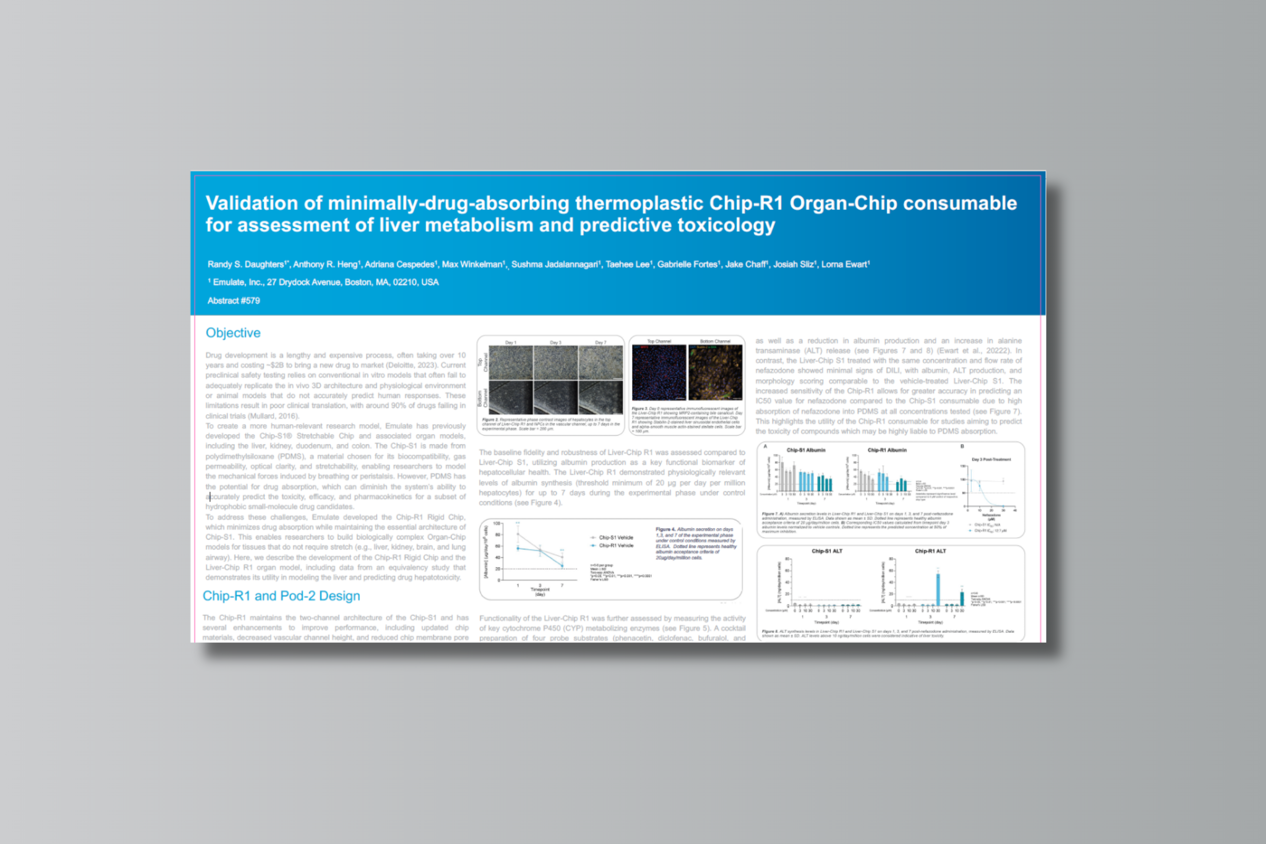
Liver-Chip R1 Application Note

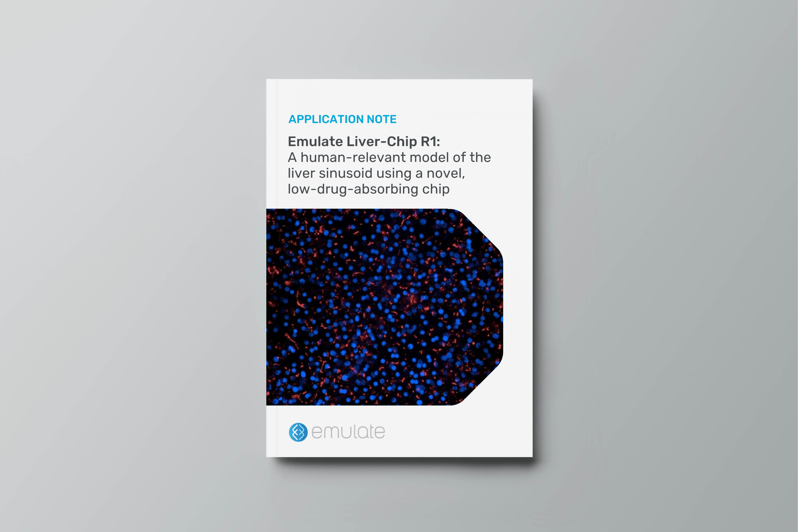
The Chip-R1 Rigid Chip minimizes drug absorption while maintaining the essential architecture of Chip-S1. This enables researchers to build biologically complex Organ-Chip models for tissues that do not require stretch (e.g., liver, kidney, brain, and lung airway).

Here, we describe the development of the Chip-R1 Rigid Chip and the Liver-Chip R1 organ model, including data from an equivalency study that demonstrates its utility in modeling the liver and predicting drug hepatotoxicity.

  • The Chip-R1™ Rigid Chip has the same two-channel configuration as the Chip-S1® Stretchable Chip, with several updates, including reduced drug absorption.
  • The Liver-Chip R1 demonstrates robust liver functionality, as indicated by morphology, marker expression, albumin production, and drug metabolism.
  • The Liver-Chip R1 displays increased sensitivity to detecting the drug-induced liver injury risk of small-molecule drugs with absorption liability in PDMS.
  • The Chip-R1 exhibits reduced absorption of a range of small molecules with diverse physicochemical properties.

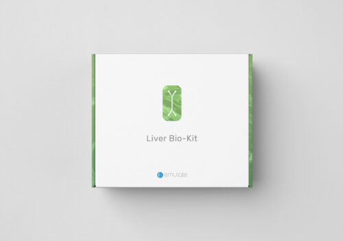
Liver-Chip R1 BioKit Data Sheet

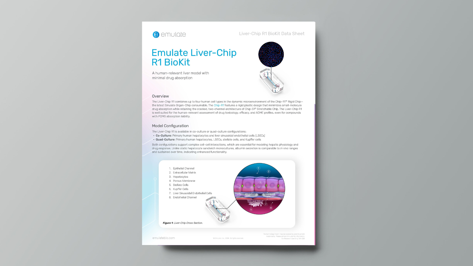
The Liver-Chip R1 BioKit contains all the components needed to build a Liver-Chip using the Chip-R1® Rigid Chip. Designed to minimize drug absorption and enhance biological modeling by using low-drug-absorbing materials, this model is particularly well-suited for applications in which drug absorption is a concern, including human-relevant assessments of drug toxicology, efficacy, and ADME profiles​​.

Human quad liver-on-chip system as a tool toward bridging the gap between animals and humans regarding toxicology and pharmacology of a cannabidiol-rich cannabis extract

Organ Model: Liver

Application: Toxicology

Abstract: Cannabidiol (CBD) is a major phytocannabinoid from Cannabis sativa. It is currently widely available and widely used in the USA, but despite its rapid progress to market, the pharmacology and toxicology of both CBD and cannabidiol-rich cannabis extracts (CRCE) remain largely unknown. The goals of this study were to investigate the potential of a novel human microphysiological system to emulate CRCE-induced hepatotoxicity and pharmacological properties demonstrated in animal models. For this purpose, C57BL6/J male mice were subjected to dosing with either 0, 61.5, 184.5, or 615 mg/kg of CRCE for 10 days. The liver-on-chip system, incorporating human primary hepatocytes, sinusoidal endothelial cells, as well as Kupffer and stellate cells was subjected to 0, 300, 1,200, or 4,400 ng/mL of CRCE (8 h exposure followed by 16 h washout) for 5 days. Administration of CRCE in mice resulted in nearly 4-fold elevations of plasma ALT at 615 mg/kg (p < 0.01) and a dose-dependent decrease in intrahepatic miR-122. Elevated levels of ALT, paralleled by decreased intrahepatic and increased effluent levels of miR-122, were also observed in the liver-on-chip, although these results were not statistically significant. Exposure to CRCE resulted in a robust and dose-dependent induction of key cytochrome P450 enzymes, namely Cyp1a2Cyp2b6 (CYP2B10), Cyp2e1, and Cyp2c9 (CYP2C19) in both mouse livers and liver-on-chip. The results of this study demonstrate the congruence between the responses observed in mouse and human liver-on-chip experimental systems and provide evidence of the potential microphysiological systems hold for translating animal data into clinical practice.

Organ chips with integrated multifunctional sensors enable continuous metabolic monitoring at controlled oxygen levels

Organ Model: Small Intestine & Liver

Application: Organ-on-a-Chip Technology

Abstract: Despite remarkable advances in Organ-on-a-chip (Organ Chip) microfluidic culture technology, recreating tissue-relevant physiological conditions, such as the region-specific oxygen concentrations, remains a formidable technical challenge, and analysis of tissue functions is commonly carried out using one analytical technique at a time. Here, we describe two-channel Organ Chip microfluidic devices fabricated from polydimethylsiloxane and gas impermeable polycarbonate materials that are integrated with multiple sensors, mounted on a printed circuit board and operated using a commercially available Organ Chip culture instrument. The novelty of this system is that it enables the recreation of physiologically relevant tissue-tissue interfaces and oxygen tension as well as non-invasive continuous measurement of transepithelial electrical resistance, oxygen concentration and pH, combined with simultaneous analysis of cellular metabolic activity (ATP/ADP ratio), cell morphology, and tissue phenotype. We demonstrate the reliable and reproducible functionality of this system in living human Gut and Liver Chip cultures. Changes in tissue barrier function and oxygen tension along with their functional and metabolic responses to chemical stimuli (e.g., calcium chelation, oligomycin) were continuously and noninvasively monitored on-chip for up to 23 days. A physiologically relevant microaerobic microenvironment that supports co-culture of human intestinal cells with living Lactococcus lactis bacteria also was demonstrated in the Gut Chip. The integration of multi-functional sensors into Organ Chips provides a robust and scalable platform for the simultaneous, continuous, and non-invasive monitoring of multiple physiological functions that can significantly enhance the comprehensive and reliable evaluation of engineered tissues in Organ Chip models in basic research, preclinical modeling, and drug development.

Developing an RNA Signature for Radiation Injury Using a Human Liver-on-a-Chip Model

Organ Model: Liver

Application: Toxicology

Abstract: Radiation exposure in a therapeutic setting or during a mass casualty event requires improved medical triaging, where the time to delivery and quantity of medical countermeasures are critical to survival. Radiation-induced liver injury (RILI) and fibrosis can lead to death, but clinical symptoms manifest late in disease pathogenesis and there is no simple diagnostic test to determine RILI. Because animal models do not completely recapitulate clinical symptoms, we used a human liver-on-a-chip model to identify biomarkers of RILI. The goals of this study were: 1. to establish a microfluidic liver-on-a-chip device as a physiologically relevant model for studying radiation-induced tissue damage; and 2. to determine acute changes in RNA expression and biological pathway regulation that identify potential biomarkers and mechanisms of RILI. To model functional human liver tissue, we used the Emulate organ-on-a-chip system to establish a co-culture of human liver sinusoidal endothelial cells (LSECs) and hepatocytes. The chips were subject to 0 Gy (sham), 1 Gy, 4 Gy, or 10 Gy irradiation and cells were collected at 6 h, 24 h, or 7 days postirradiation for RNA isolation. To identify significant expression changes in messenger RNA (mRNA) and long non-coding RNA (lncRNA), we performed RNA sequencing (RNASeq) to conduct whole transcriptome analysis. We found distinct differences in expression patterns by time, dose, and cell type, with higher doses of radiation resulting in the most pronounced expression changes, as anticipated. Ingenuity Pathway Analysis indicated significant inhibition of the cell viability pathway 24 h after 10 Gy exposure in LSECs but activation of this pathway in hepatocytes, highlighting differences between cell types despite receiving the same radiation dose. Overall, hepatocytes showed fewer gene expression changes in response to radiation, with only 3 statistically significant differentially expressed genes at 7 days: APOBEC3H, PTCHD4, and GDNF. We further highlight lncRNA of interest including DINO and PURPL in hepatocytes and TMPO-AS1 and PRC-AS1 in LSECs, identifying potential biomarkers of RILI. We demonstrated the potential utility of a human liver-on-a-chip model with primary cells to model organ-specific radiation injury, establishing a model for radiation medical countermeasure development and further biomarker validation. Furthermore, we identified biomarkers that differentiate radiation dose and defined cell-specific targets for potential radiation mitigation therapies.

Evaluating the Hepatotoxicity of Cannabidiol, Cannabinol, Cannabichromene and Cannabigerol Using a Human Quad-Culture Liver-Chip

Abstract

As the popularity of hemp-derived products grows, understanding the prospective hepatotoxicity of cannabinoids becomes crucial for personal safety. Despite conflicting evidence from limited human clinical studies, a safety gap persists, and no standard threshold has been established for these various cannabinoid-containing products. Furthermore, the hepatotoxicity potential of other cannabinoids like Cannabinol (CBN), Cannabichromene (CBC), and Cannabigerol (CBG) remains largely unexplored.

The current study was designed to address some of these gaps by utilizing microphysiological systems (MPS), such as the Emulate Quad-Culture Liver-Chip, which adhere to IQ MPS consortia guidelines, as an alternative to animal testing. Here, pressure-driven flow for compound delivery was used with primary human cells in an in vivo-like arrangement, separated by a semi-porous membrane to allow for crosstalk between the hepatocytes and nonparenchymal cells (NPCs). Hepatotoxicity was compared among CBD, CBN, CBC, and CBG, in parallel with the known hepatotoxic compound acetaminophen (APAP), in a three-point concentration response evaluation (0.24, 3, or 4.7 µM). The assessment encompassed morphological effects, hepatocyte function, and potential mechanisms of action over a 7-day continuous dosing period. These evaluations included live imaging for mitochondrial dysfunction, total reactive oxygen species (ROS), and inflammatory cytokines derived from effluent-based sampling in the Emulate platform. 

Morphological analysis revealed 3 or 4.7 µM CBD impacting hepatocytes by Day 7, while CBG exhibited no visible changes compared to the control. Endpoints evaluating hepatocyte function and viability indicated that LDH release increased only with 4.7 µM CBD, CBN, and CBC. CBD, CBN, and CBG did not significantly affect albumin, ALT, or AST, while 4.7 µM CBC significantly decreased albumin production. Inflammatory cytokines increased at high concentrations of CBD, CBC, and CBG. ROS and mitochondrial function displayed different responses among the cannabinoids affecting the NPCs more versus the hepatocytes. Up until now, the majority of what is known regarding these compounds is mostly murine-based. Hence, this study was an effort to leverage the use of advanced, human-relevant MPS to carefully assess and compare the hepatotoxicity of CBD and other cannabinoids, shedding light on their safety in foods and health products.

Organ-on-a-chip for studying immune cell adhesion to liver sinusoidal endothelial cells: the potential for testing immunotherapies and cell therapy trafficking

Organ Model: Liver

Application: Immunology & Inflammation

Abstract: Immunotherapy has changed the landscape of treatment options for patients with hepatocellular cancer. Checkpoint inhibitors are now standard of care for patients with advanced tumours, yet the majority remain resistant to this therapy and urgent approaches are needed to boost the efficacy of these agents. Targeting the liver endothelial cells, as the orchestrators of immune cell recruitment, within the tumour microenvironment of this highly vascular cancer could potentially boost immune cell infiltration. We demonstrate the successful culture of primary human liver endothelial cells in organ-on-a-chip technology followed by perfusion of peripheral blood mononuclear cells. We confirm, with confocal and multiphoton imaging, the capture and adhesion of immune cells in response to pro-inflammatory cytokines in this model. This multicellular platform sets the foundation for testing the efficacy of new therapies in promoting leukocyte infiltration across liver endothelium as well as a model for testing cell therapy, such as chimeric antigen receptor (CAR)-T cell, capture and migration across human liver endothelium.

Beyond the hype and toward application: liver complex in vitro models in preclinical drug safety

Overview

Drug induced Liver-Injury (DILI) is a leading cause of drug attrition, and complex in vitro models (CIVMs)—including three dimensional (3D) spheroids, Organ-Chips, 3D bio printed tissues, and flow-based systems—could improve preclinical prediction. Although CIVMs have demonstrated good sensitivity and specificity, in DILI detection their adoption remains limited.

This article describes DILI, the challenges with its prediction and the current strategies, and the models that are being used to study it. It reviews data from industry-FDA collaborations and strategic partnerships and finishes with an outlook of CIVMs in preclinical toxicity testing.

On the potential of the Human Liver-Chip as a model of cholestatic toxicity

Abstract

Bile acids are an essential component of bile, which aids digestion in the form of emulsification and absorption of lipids. Primary bile acids (cholic and chenodeoxycholic acid) are synthesized in the liver, can be conjugated with glycine or taurine, concentrated in the gall bladder, and circulated to the intestine during digestion. Primary bile acids are converted to secondary bile acids (deoxycholic and lithocholic acid) by intestinal bacteria and are recirculated back to the liver via enterohepatic circulation. In a healthy liver, bile acids are secreted from hepatocytes into bile canaliculi through the bile salt export pump (BSEP) which is involved in maintaining bile acid homeostasis. When bile flow is disrupted, increasing levels of intrahepatic bile acids cause cholestatic injury. Cholestasis can be induced by drugs like troglitazone (TROG), a diabetes therapy that was discontinued after causing liver toxicity in clinical trials partly via inhibition of BSEP. To establish a physiologically relevant model of drug-induced cholestasis, Emulate’s human Quad-Culture Liver-Chip was incubated with bile acids in the presence or absence of TROG and monitored for signs of toxicity and BSEP inhibition. A mixture of glycine-conjugated primary and secondary bile acids, including glycocholic acid (GCA), glycodeoxycholic acid (GDCA) and glycochenodeoxycholic acid (GCDCA), were chosen as they are three of the most abundant bile acids in human serum (comprising ~68% of the
total population) and were administered at concentrations ranging from 1-5X human Cmax.

Evaluating the immunotoxicity of CD137-induced agonism on the Emulate Human Liver-Chip

Abstract

The field of cancer immunotherapy is rapidly growing but is challenged by our inability to predict treatment efficacy, patient response and adverse effects. Agonistic antibodies toward T cell co-stimulatory molecules like Urelumab (targeting CD137) are one class of immunotherapy that has shown unparalleled efficacy in murine models of cancer. However, in clinical trials (NCT00309023), Urelumab doses ≥ 1 mg/kg resulted in significant hepatotoxicity (mainly transaminitis), suggesting murine and NHP preclinical assessment failed to predict this clinical safety outcome. While the mechanism of clinical liver toxicity has not been fully elucidated, studies with humanized mice (with human hematopoietic cells) indicate immune-mediated DILI (drug-induced liver injury). Therefore, to model hepatotoxicity associated with Urelumab, we created a novel, immuno-competent human Quad-Culture Liver-Chip using human peripheral blood mononuclear cells (PBMCs) that contain lymphocytes, NK cells and CD137+ monocytes to drive immunotoxicity.