A Human Proximal Tubule Kidney-Chip for Accelerated Therapeutic Development

Abstract

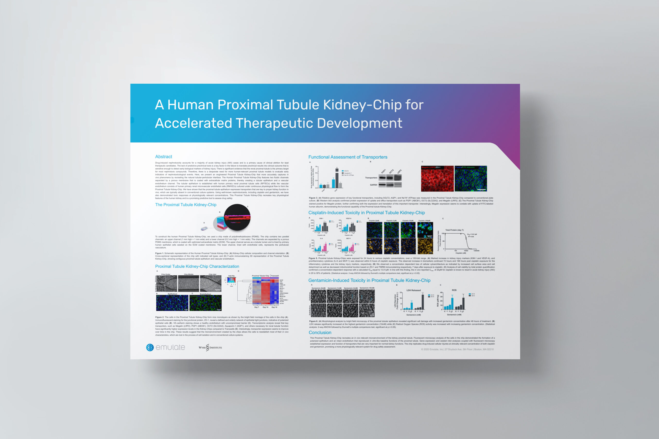
Drug-induced nephrotoxicity accounts for a majority of acute kidney injury (AKI) cases and is a primary cause of clinical attrition for lead therapeutic candidates. The lack of predictive preclinical tools is a key factor in the failure to translate preclinical results into clinical outcome that is sensitive enough to detect early biological markers of kidney injury. There is significant evidence that the renal proximal tubule is the primary target for most nephrotoxic compounds. Therefore, there is a desperate need for more human-relevant proximal tubule models to evaluate early indicators of nephrotoxicological events. Here, we present an engineered Proximal Tubule Kidney-Chip that more accurately captures in vivo phenomena by recreating the natural tubular-peritubular interface. The Human Proximal Tubule Kidney-Chip features two fluidic channels separated by a porous membrane that is coated with extracellular matrix proteins, thereby creating a tubular epithelium and a vascular endothelium channel. The tubular epithelium is established with human primary renal proximal tubule cells (RPTECs) while the vascular endothelium consists of human primary renal microvascular endothelial cells (RMVECs) cultured under continuous physiological flow to form the Proximal Tubule Kidney-Chip. We have shown that the proximal tubule epithelium expresses transporters that are key to proper kidney function in vivo, which are typically absent in conventional culture systems. Using well-known nephrotoxicants, including cisplatin and gentamicin, we have also demonstrated toxic responses at physiologically relevant concentrations. This Proximal Tubule Kidney-Chip recreates key physiological features of the human kidney and is a promising predictive tool to assess drug safety.

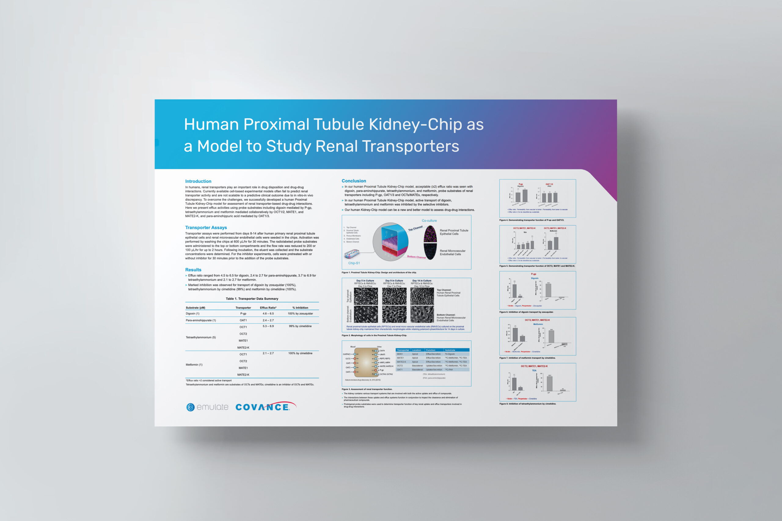
Human Proximal Tubule Kidney-Chip as a Model to Study Renal Transporters

Introduction

In humans, renal transporters play an important role in drug disposition and drug-drug interactions. Currently available cell-based experimental models often fail to predict renal transporter activity and are not scalable to a predictive clinical outcome due to in vitro-in vivo discrepancy. To overcome the challenges, we successfully developed a human Proximal Tubule Kidney-Chip model for assessment of renal transporter-based drug-drug interactions. Here we present efflux activities using probe substrates including digoxin mediated by P-gp, tetraethylammonium and metformin mediated collaboratively by OCT1/2, MATE1, and MATE2-K, and para-aminohippuric acid mediated by OAT1/3.

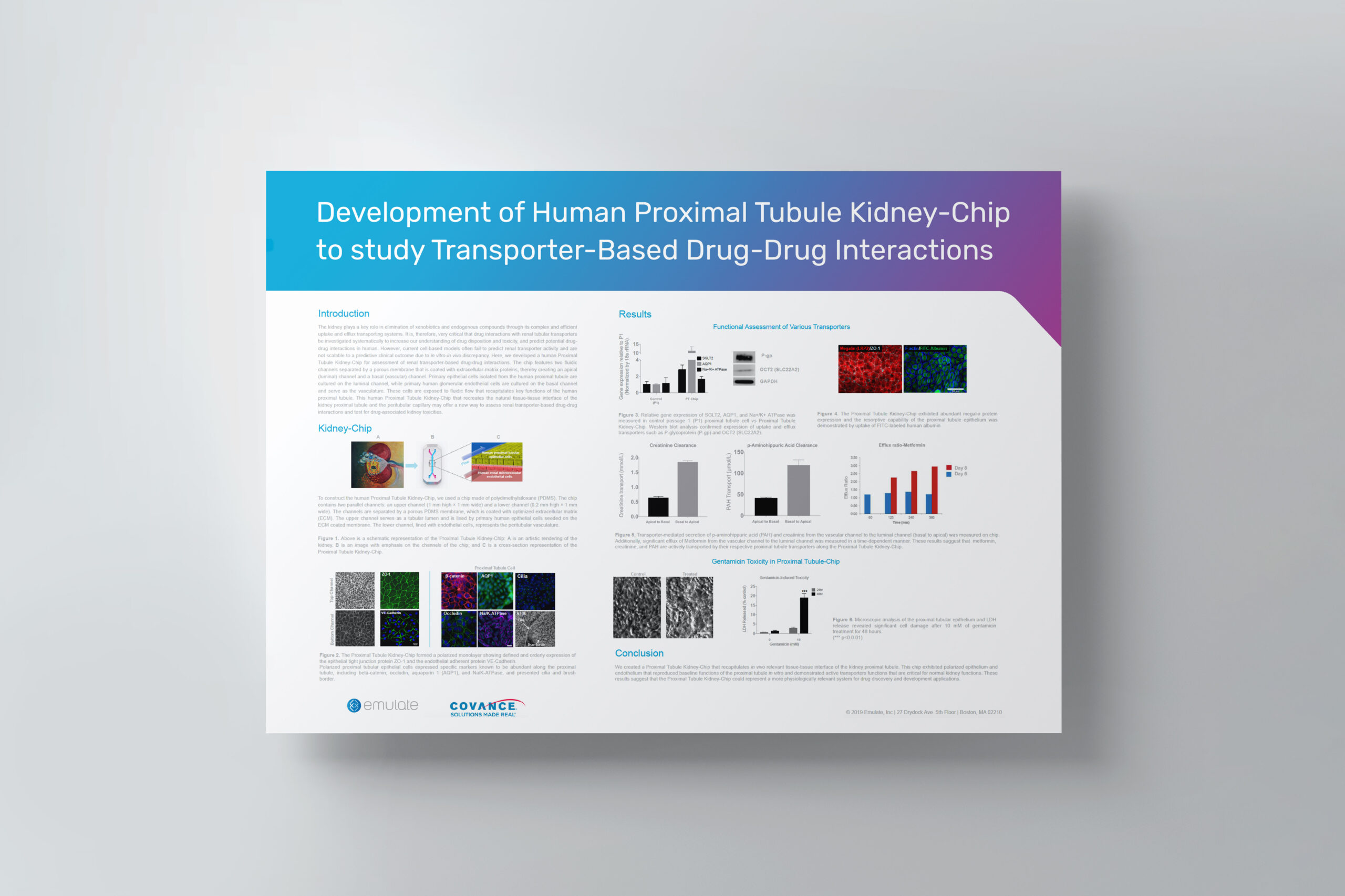
Development of Human Proximal Tubule Kidney-Chip to Study Transporter-Based Drug-Drug Interactions

Abstract

The kidney plays a key role in elimination of xenobiotics and endogenous compounds through its complex and efficient uptake and efflux transporting systems. It is, therefore, very critical that drug interactions with renal tubular transporters be investigated systematically to increase our understanding of drug disposition and toxicity, and predict potential drug-drug interactions in humans. However, current cell-based models often fail to predict renal transporter activity and are not scalable to a predictive clinical outcome due to in vitroin vivo discrepancy. Here, we developed a human Proximal Tubule Kidney-Chip for assessment of renal transporter-based drug-drug interactions. The chip features two fluidic channels separated by a porous membrane that is coated with extracellular-matrix proteins, thereby creating an apical (luminal) channel and a basal (vascular) channel. Primary epithelial cells isolated from the human proximal tubule are cultured on the luminal channel, while primary human glomerular endothelial cells are cultured on the basal channel and serve as the vasculature. These cells are exposed to fluidic flow that recapitulates key functions of the human proximal tubule. This human Proximal Tubule Kidney-Chip that recreates the natural tissue-tissue interface of the kidney proximal tubule and the peritubular capillary may offer a new way to assess renal transporter-based drug-drug interactions and test for drug-associated kidney toxicities.

REST and Stress Resistance in the Ageing Kidney

Organ Model: Kidney (Glomerulus mouse)

Application: Model Development

Abstract: CKD is associated with the loss of functional nephrons, leading to increased mechanical and metabolic stress in the remaining cells, particularly for cells constituting the filtration barrier, such as podocytes. The failure of podocytes to mount an adequate stress response can lead to further nephron loss and disease progression. However, the mechanisms that regulate this degenerative process in the kidney are unknown.

Evaluating the Kidney-Chip for Transporter-Mediated Drug-Drug Interactions Applications

Webinar Overview

In this on-demand webinar, Covance and Emulate present data on the characterization of the Kidney-Chip as a model to study and predict potential drug-drug interactions.

In the pharmaceutical industry, there is a need for more human-relevant preclinical models that can enable a mechanistic understanding of human diseases and drug action. 

Organ-Chips can help address this need by recreating in vivo-relevant function and key disease phenotypes in a more physiological microenvironment than conventional cell-based models. Covance and Emulate are collaborating to evaluate the Proximal Tubule Kidney-Chip to study transporter-mediated DDIs to fill the gap between current in vitro options and clinical trials.

In this webinar, scientists from Emulate and Covance showcase promising new data on characterization of the Kidney-Chip as a model to study and predict potential drug-drug interactions.


Featured Presenters

Steve Anderson, PhD
CSO, Covance

Steve outlines the need for more human-relevant cell-based systems to improve translation of preclinical data to the clinic and get safer, more efficacious therapeutics to patients.

Kyung-Jin (KJ) Jang, PhD
VP of Technology Implementation & Field Science, Emulate

KJ highlights the Human Emulation System, an Organs-on-Chips platform that recreates the required microenvironment to enable physiologically relevant function in a human cell-based system, as a new solution to study and predict transporter-mediated DDIs.

Donald McKenzie, PhD
Global Business Lead for Drug Metabolism & Lead Optimization, Covance

Donald presents proof-of-concept data to characterize the Proximal Tubule Kidney-Chip including efflux activities using probe substrates such as digoxin mediated by P-gp; tetraethylammonium and metformin mediated collaboratively by OCT1/2, MATE1, and MATE2-K; and para aminohippuric acid mediated by OAT1/3. The data also includes functional modulation of efflux by known inhibitors of specific transporters.


Directed differentiation of human induced pluripotent stem cells into mature kidney podocytes and establishment of a Glomerulus Chip

Organ Model: Kidney (Glomerulus)

Applications: Model development, ADME, Toxicology

Abstract: Protocols have been established to direct the differentiation of human induced pluripotent stem (iPS) cells into nephron progenitor cells and organoids containing many types of kidney cells, but it has been difficult to direct the differentiation of iPS cells to form specific types of mature human kidney cells with high yield. Here, we describe a detailed protocol for the directed differentiation of human iPS cells into mature, post-mitotic kidney glomerular podocytes with high (>90%) efficiency within 26 d and under chemically defined conditions, without genetic manipulations or subpopulation selection. We also describe how these iPS cell-derived podocytes may be induced to form within a microfluidic organ-on-a-chip (Organ Chip) culture device to build a human kidney Glomerulus Chip that mimics the structure and function of the kidney glomerular capillary wall in vitro within 35 d (starting with undifferentiated iPS cells). The podocyte differentiation protocol requires skills for culturing iPS cells, and the development of a Glomerulus Chip requires some experience with building and operating microfluidic cell culture systems. This method could be useful for applications in nephrotoxicity screening, therapeutic development, and regenerative medicine, as well as mechanistic study of kidney development and disease.

Mature induced-pluripotent-stem-cell-derived human podocytes reconstitute kidney glomerular-capillary-wall function on a chip

Organ Model: Kidney (Glomerulus)

Applications: Model development, ADME, Toxicology

Abstract: An in vitro model of the human kidney glomerulus – the major site of blood filtration – could facilitate drug discovery and illuminate kidney-disease mechanisms. Microfluidic organ-on-a-chip technology has been used to model the human proximal tubule, yet a kidney-glomerulus-on-a-chip has not been possible because of the lack of functional human podocytes – the cells that regulate selective permeability in the glomerulus. Here, we demonstrate an efficient (> 90%) and chemically defined method for directing the differentiation of human induced pluripotent stem (hiPS) cells into podocytes that express markers of the mature phenotype (nephrin+, WT1+, podocin+, Pax2-) and that exhibit primary and secondary foot processes. We also show that the hiPS-cell-derived podocytes produce glomerular basement-membrane collagen and recapitulate the natural tissue/tissue interface of the glomerulus, as well as the differential clearance of albumin and inulin, when co-cultured with human glomerular endothelial cells in an organ-on-a-chip microfluidic device. The glomerulus-on-a-chip also mimics adriamycin-induced albuminuria and podocyte injury. This in vitro model of human glomerular function with mature human podocytes may facilitate drug development and personalized-medicine applications.

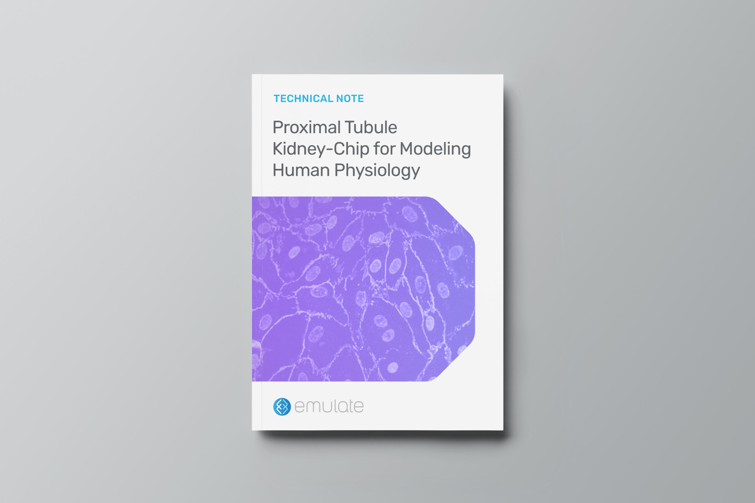
Proximal Tubule Kidney-Chip for Modeling Human Physiology

Overview

In the pharmaceutical industry, there is a need for more human-relevant preclinical models to improve translation to clinical trials. Organ-Chips can help address this need by recreating in vivo-relevant function in a more physiological microenvironment than conventional cell-based models. In this technical note, we review how our Chip-S1 can be used to create a comprehensive recapitulation of the proximal tubule

Key highlights:

  • Kidney-Chip emulates in vivo-like physiology by incorporating a co-culture of primary human kidney-specific epithelial and endothelial cells in a dynamic microenvironment.
  • Continuous perfusion of media ensures cells are exposed to sufficient amounts of test compounds, metabolites, or other stimulus factors
  • Cells can be fixed and imaged for further analysis or removed and processed for genomic studies
  • Potential applications include investigate of drug candidate toxicity as well as evaluation of transporter-mediated drug-drug interactions.

Human kidney proximal tubule-on-a-chip for drug transport and nephrotoxicity assessment

Organ Model: Kidney (Proximal Tubule)

Application: ADME & Toxicology

Abstract: Kidney toxicity is one of the most frequent adverse events reported during drug development. The lack of accurate predictive cell culture models and the unreliability of animal studies have created a need for better approaches to recapitulate kidney function in vitro. Here, we describe a microfluidic device lined by living human kidney epithelial cells exposed to fluidic flow that mimics key functions of the human kidney proximal tubule. Primary kidney epithelial cells isolated from human proximal tubule are cultured on the upper surface of an extracellular matrix-coated, porous, polyester membrane that splits the main channel of the device into two adjacent channels, thereby creating an apical ‘luminal’ channel and a basal ‘interstitial’ space. Exposure of the epithelial monolayer to an apical fluid shear stress (0.2 dyne cm(-2)) that mimics that found in living kidney tubules results in enhanced epithelial cell polarization and primary cilia formation compared to traditional Transwell culture systems. The cells also exhibited significantly greater albumin transport, glucose reabsorption, and brush border alkaline phosphatase activity. Importantly, cisplatin toxicity and Pgp efflux transporter activity measured on-chip more closely mimic the in vivo responses than results obtained with cells maintained under conventional culture conditions. While past studies have analyzed kidney tubular cells cultured under flow conditions in vitro, this is the first report of a toxicity study using primary human kidney proximal tubular epithelial cells in a microfluidic ‘organ-on-a-chip’ microdevice. The in vivo-like pathophysiology observed in this system suggests that it might serve as a useful tool for evaluating human-relevant renal toxicity in preclinical safety studies.