Emulate BioKits: Innovative Case Studies in Disease Modeling and Immunotherapy Safety

In this webinar, Emeli Chatterjee, Postdoctoral Researcher at Massachusetts General Hospital, and Ben Swenor, Senior Science Liaison at Emulate, present case studies from three peer-reviewed papers highlighting how Organ-Chips have been used to study cardiorenal syndrome, environmental enteric dysfunction (EED), and immunotherapy safety. View now to learn how Organ-Chips can be used to investigate complex disease mechanisms and evaluate immunotherapy safety.

Case study topics include: 

  • Cardiorenal Syndrome: Investigating the role of extracellular vesicles from patients with cardiorenal syndrome on renal injury using the Kidney-Chip, offering insights into disease mechanisms and potential therapeutic targets. 
  • Immuno-oncology Safety: Using the Colon Intestine-Chip and Duodenum Intestine-Chip to evaluate the on-target, off-tumor safety of T-cell bispecific antibodies. 
  • Environmental enteric dysfunction (EED): Replicating EED disease mechanisms with the Duodenum Intestine-Chip, comparing the effects of malnutrition with healthy vs. patient-derived tissue to uncover novel therapeutic targets. 

Read the author Q&A here.

Engineered human iPS cell models reveal altered podocytogenesis and glomerular capillary wall in CHD-associated SMAD2 mutations 

Organ Model: Kidney (Glomerulus)

Application: Model Development

Abstract: Early developmental programming involves extensive cell lineage diversification through shared molecular signaling networks. Clinical observations of congenital heart disease (CHD) patients carrying SMAD2 genetic variants revealed correlations with multi-organ impairments at the developmental and functional levels. For example, many CHD patients present with glomerulosclerosis, periglomerular fibrosis, and albuminuria. Still, it remains largely unknown whether SMAD2 variants associated with CHD can directly alter kidney cell fate, tissue patterning, and organ-level function. To address this question, we engineered human iPS cells (iPSCs) and organ-on-a-chip systems to uncover the role of pathogenic SMAD2 variants in kidney podocytogenesis. Our results show that abrogation of SMAD2 causes altered patterning of the mesoderm and intermediate mesoderm (IM) cell lineages, which give rise to nearly all kidney cell types. Upon further differentiation of IM cells, the mutant podocytes failed to develop arborizations and interdigitations. A reconstituted glomerulus-on-a-chip platform exhibited significant proteinuria as clinically observed in glomerulopathies. This study implicates CHD-associated SMAD2 mutations in kidney tissue malformation and provides opportunities for therapeutic discovery in the future.

An ultrathin membrane mediates tissue-specific morphogenesis and barrier function in a human kidney chip

Organ Model: Kidney (Glomerulus)

Application: Model Development

Abstract: Organ-on-chip (OOC) systems are revolutionizing tissue engineering by providing dynamic models of tissue structure, organ-level function, and disease phenotypes using human cells. However, nonbiological components of OOC devices often limit the recapitulation of in vivo–like tissue-tissue cross-talk and morphogenesis. Here, we engineered a kidney glomerulus-on-a-chip that recapitulates glomerular morphogenesis and barrier function using a biomimetic ultrathin membrane and human-induced pluripotent stem cells. The resulting chip comprised a proximate epithelial-endothelial tissue interface, which reconstituted the selective molecular filtration function of healthy and diseased kidneys. In addition, fenestrated endothelium was successfully induced from human pluripotent stem cells in an OOC device, through in vivo–like paracrine signaling across the ultrathin membrane. Thus, this device provides a dynamic tissue engineering platform for modeling human kidney–specific morphogenesis and function, enabling mechanistic studies of stem cell differentiation, organ physiology, and pathophysiology.

Investigating a Functional Role for EVs in Modulating Disease Signaling Pathways in Organ-on-Chip Models

Webinar Abstract

Featured session at Boston MPS Day, which took place on 11/1/2023.

Physiological Replication of the Human Glomerulus Using a Triple Culture Microphysiological System

Organ Model: Kidney (Glomerulus)

Application: Model Development

Abstract: The function of the glomerulus depends on the complex cell-cell/matrix interactions and replication of this in vitro would aid biological understanding in both health and disease. Previous models do not fully reflect all cell types and interactions present as they overlook mesangial cells within their 3D matrix. Herein, the development of a microphysiological system that contains all resident renal cell types in an anatomically relevant manner is presented. A detailed transcriptomic analysis of the contributing biology of each cell type, as well as functionally appropriate albumin retention in the system, is demonstrated. The important role of mesangial cells is shown in promoting the health and maturity of the other cell types. Additionally, a comparison of the incremental advances that each individual cell type brings to the phenotype of the others demonstrates that glomerular cells in simple 2D culture exhibit a state more reflective of the dysfunction observed in human disease than previously recognized. This in vitro model will expand the capability to investigate glomerular biology in a more translatable manner by the inclusion of the important mesangial cell compartment.

Circulating extracellular vesicles in human cardiorenal syndrome promote renal injury in Kidney on Chip system

Organ Model: Kidney (Proximal Tubule)

Application: Inflammation

Abstract: Background: Cardiorenal syndrome (CRS) – renal injury during heart failure (HF) – is linked to high morbidity. Whether circulating extracellular vesicles (EVs) and their RNA cargo directly impact its pathogenesis remains unclear. Methods: We investigated the role of circulating EVs from patients with CRS on renal epithelial/endothelial cells using a microfluidic kidney-on-chip (KOC) model. The small RNA cargo of circulating EVs was regressed against serum creatinine to prioritize subsets of functionally relevant EV-miRNAs and their mRNA targets investigated using in silico pathway analysis, human genetics, and interrogation of expression in the KOC model and in renal tissue. The functional effects of EV-RNAs on kidney epithelial cells were experimentally validated. Results: Renal epithelial and endothelial cells in the KOC model exhibited uptake of EVs from patients with HF. HF-CRS EVs led to higher expression of renal injury markers (IL18, LCN2, HAVCR1) relative to non-CRS EVs. A total of 15 EV-miRNAs were associated with creatinine, targeting 1,143 gene targets specifying pathways relevant to renal injury, including TGF-? and AMPK signaling. We observed directionally consistent changes in the expression of TGF-? pathway members (BMP6, FST, TIMP3) in the KOC model exposed to CRS EVs, which were validated in epithelial cells treated with corresponding inhibitors and mimics of miRNAs. A similar trend was observed in renal tissue with kidney injury. Mendelian randomization suggested a role for FST in renal function. Conclusion: Plasma EVs in patients with CRS elicit adverse transcriptional and phenotypic responses in a KOC model by regulating biologically relevant pathways, suggesting a role for EVs in CRS.

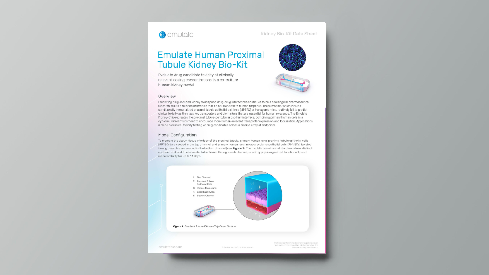
Proximal Tubule Kidney-Chip S1 BioKit Data Sheet

OVERVIEW

The Proximal Tubule Kidney-Chip S1 BioKit includes the essential components needed to create the Proximal Tubule Kidney-Chip—including Emulate qualified cells—and is available in multiple sizes to meet various study needs.

Assessing Nephrotoxicity and Drug-Drug Interactions with the Emulate Proximal Tubule Kidney-Chip

Webinar Abstract

Predicting drug-induced kidney toxicity and drug-drug interactions during preclinical development continues to be challenging due to a researchers’ reliance on immortalized cell lines and animal models that do not translate to human response. These translational issues have real-world impacts in the clinic, with nephrotoxicity causing preclinical attrition and an estimated 19% of failures during phase 3 clinical trials1

The Emulate Proximal Tubule Kidney-Chip has been developed to address these challenges, enabling researchers to more accurately model nephrotoxicity and drug-drug interactions to improve clinical success. By incorporating primary human kidney epithelial cells with tissue-specific endothelium in a dynamic microenvironment, the Proximal Tubule Kidney-Chip achieves a proper kidney phenotype exhibiting normal epithelial cell polarity and morphology and demonstrating in vivo-relevant functional transporter activity. 

View this data-driven webinar to learn how you can use Organ-on-a-Chip technology to overcome the limitations of conventional kidney models and gain more confidence before taking your drug candidate into the clinic. 

Key Advantages of the Proximal Tubule Kidney-Chip:   

  • Retains characteristic kidney functionality for up to 14 days 
  • Improved cytoarchitecture, polarization, and kidney marker expression due to a dynamic microenvironment  
  • Improved functionality of key renal transporters (Megalin, Cubulin, P-gp, MATE1, MATE2-K, OAT1, OAT3, and OCT2, and more)  
  • Demonstrated ability to model drug-induced nephrotoxicity and renal transporter-mediated drug-drug interactions  

A Personalized Glomerulus Chip Engineered from Stem Cell-Derived Epithelium and Vascular Endothelium

Organ Model: Kidney (Glomerulus)

Application: Toxicology

Abstract: Progress in understanding kidney disease mechanisms and the development of targeted therapeutics have been limited by the lack of functional in vitro models that can closely recapitulate human physiological responses. Organ Chip (or organ-on-a-chip) microfluidic devices provide unique opportunities to overcome some of these challenges given their ability to model the structure and function of tissues and organs in vitro. Previously established organ chip models typically consist of heterogenous cell populations sourced from multiple donors, limiting their applications in patient-specific disease modeling and personalized medicine. In this study, we engineered a personalized glomerulus chip system reconstituted from human induced pluripotent stem (iPS) cell-derived vascular endothelial cells (ECs) and podocytes from a single patient. Our stem cell-derived kidney glomerulus chip successfully mimics the structure and some essential functions of the glomerular filtration barrier. We further modeled glomerular injury in our tissue chips by administering a clinically relevant dose of the chemotherapy drug Adriamycin. The drug disrupts the structural integrity of the endothelium and the podocyte tissue layers, leading to significant albuminuria as observed in patients with glomerulopathies. We anticipate that the personalized glomerulus chip model established in this report could help advance future studies of kidney disease mechanisms and the discovery of personalized therapies. Given the remarkable ability of human iPS cells to differentiate into almost any cell type, this work also provides a blueprint for the establishment of more personalized organ chip and ‘body-on-a-chip’ models in the future.

Dynamic persistence of UPEC intracellular bacterial communities in a human bladder-chip model of urinary tract infection

Organ Model: Bladder

Application: Infectious Disease

Abstract: Uropathogenic Escherichia coli (UPEC) proliferate within superficial bladder umbrella cells to form intracellular bacterial communities (IBCs) during early stages of urinary tract infections. However, the dynamic responses of IBCs to host stresses and antibiotic therapy are difficult to assess in situ. We develop a human bladder-chip model wherein umbrella cells and bladder microvascular endothelial cells are co-cultured under flow in urine and nutritive media respectively, and bladder filling and voiding mimicked mechanically by application and release of linear strain. Using time-lapse microscopy, we show that rapid recruitment of neutrophils from the vascular channel to sites of infection leads to swarm and neutrophil extracellular trap formation but does not prevent IBC formation. Subsequently, we tracked bacterial growth dynamics in individual IBCs through two cycles of antibiotic administration interspersed with recovery periods which revealed that the elimination of bacteria within IBCs by the antibiotic was delayed, and in some instances, did not occur at all. During the recovery period, rapid proliferation in a significant fraction of IBCs reseeded new foci of infection through bacterial shedding and host cell exfoliation. These insights reinforce a dynamic role for IBCs as harbors of bacterial persistence, with significant consequences for non-compliance with antibiotic regimens.