

Emulate Community Publications Digest: Summer 2026 Issue

With over 150 peer-reviewed publications across 30+ organ models, Emulate Organ-Chips are empowering researchers to make game-changing scientific breakthroughs! Download this digest to easily explore all publications related to your field of research, or to just learn more about how the technology itself is developing.

New this quarter:

Fallopian Tube

  • Human fallopian tube-on-a-chip for preclinical testing of non-hormonal contraceptives with living human sperm

Intestine

  • Probiotic intervention mitigates radiation-induced intestinal injury by alleviating oxidative stress in a human gut-on-a-chip

Lung (Airway)

  • Development of an acute inhalation toxicity testing method based on a lung-on-a-chip

Lung (Alveolus)

  • Enhanced lung delivery of an immunostimulatory duplex RNA augments the antitumor activity by reshaping systemic cytokine pharmacodynamics

Lung (Alveolus)

  • Alveolus-on-a-Chip: A Novel Tool for Modeling Lung Transplant Cold Storage Ischemia/Reperfusion Injury

Placenta

  • Simvastatin Restores Uteroplacental Hemodynamics and Trophoblast Function in Obstetric Antiphospholipid Syndrome in a Placenta-on-a-Chip Model

REVIEW ARTICLES:

Female Reproductive Tract

  • Female Reproductive Tract Organ-on-Chips: Modeling Barrier Function and Drug Transport

Drug Development

  • Human organ-on-a-chip technology as a catalyst for drug discovery

PUBLISHED FROM PRE-PRINT:

Intestine (Colon)

  • Human inflammatory bowel disease-on-a-chip for modelling disease progression, cancer initiation and sex-specific effects

Lymph Node

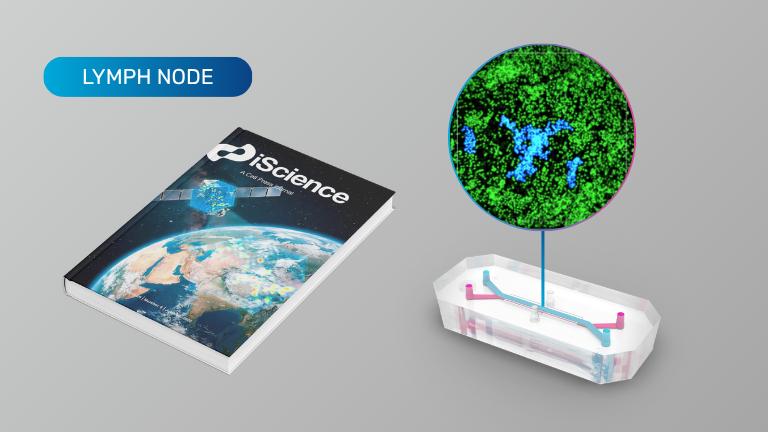
  • In vitro recapitulation of intramuscular mRNA vaccination with naive and recall antigens using a human lymphoid follicle chip platform

Vasculature

  • Human coronary artery organ-chip with circulating immune cells recapitulates anti-inflammatory effect of pulsatile wall strain
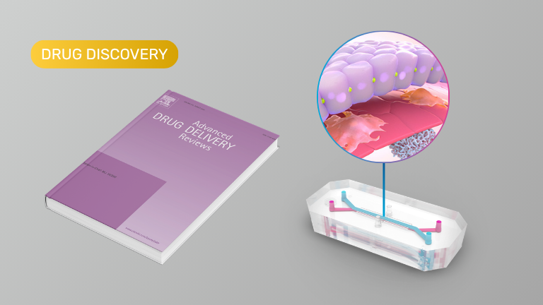
Human organ-on-a-chip technology as a catalyst for drug discovery (Review)

Application: Drug Discovery

This review highlights Organ-Chips as a more human-relevant preclinical platform that recreates key aspects of organ physiology—including tissue-tissue interfaces, dynamic fluid flow, mechanical forces, and immune interactions—to improve prediction of human drug responses. Unlike conventional cell cultures or animal models, Organ-Chips enable therapeutics to be evaluated using clinically relevant dosing routes and pharmacokinetic profiles, supporting more accurate assessment of efficacy, toxicity, and mechanisms of resistance. The authors also emphasize that combining Organ-Chips with high-throughput screening, functional genomics, and artificial intelligence could accelerate target identification and drug discovery while helping reduce the high rate of clinical trial failures. Although challenges remain around standardization and scalability, the review concludes that Organ-Chips are poised to become an increasingly important tool for drug development and regulatory safety assessment.

In vitro recapitulation of intramuscular mRNA vaccination with naive and recall antigens using a human lymphoid follicle chip platform

Organ Model: Lymph Node

Application: Immunology (vaccine development)

In this study, Organ-Chips were used to model the human immune response to intramuscular mRNA vaccines by combining a biomimetic muscle module with a microfluidic human lymphoid follicle chip (LF Chip). mRNA vaccines were administered into the muscle module containing human myoblasts and antigen-presenting cells (APCs), and the resulting APCs and soluble factors were transferred to the LF Chip to mimic lymphatic drainage, leading to antigen-specific antibody production and cytokine release. This platform enabled evaluation of both primary and recall immune responses, including neutralizing antibody generation and somatic hypermutation.

Products Used In This Publication

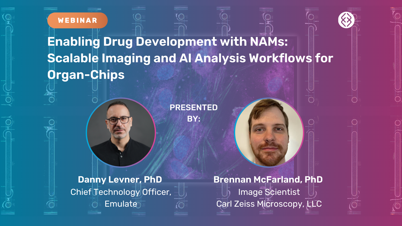
Enabling Drug Development with NAMs: Scalable Imaging and AI Analysis Workflows for Organ-Chips

Synopsis

Organ-on-a-Chip technology is emerging as a powerful New Approach Methodology (NAM) for drug development, driven by the need for more human-relevant and scalable experimental models. As these systems move toward broader adoption, a key challenge remains: generating consistent, interpretable data that supports confident experimental and translational decision-making.

This webinar examines how imaging and AI-driven analysis workflows enable Organ-Chip studies to scale from innovation to routine application. Speakers begin with an overview of Organ-on-a-Chip technology and its role in addressing translational gaps in drug discovery, highlighting how Liver-Chips are being evaluated in collaboration with regulatory agencies for better prediction of drug-induced liver injury.

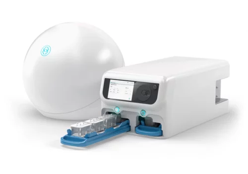
The session then explores how the newly released AVA™ Emulation System enables scalable Organ-Chip experimentation through an integrated system for incubation, microfluidic delivery, and routine imaging. Paired with AI-driven analysis, brightfield image data can be used to automate quality control by monitoring chip health, morphology, and assay performance over time across large studies.

To complete the workflow, post-study high-resolution imaging is applied to evaluate more complex biological markers, including toxicology-relevant endpoints and drug uptake. These datasets are paired with advanced analysis techniques that translate imaging data into quantitative, biologically meaningful insights.

Attendees will gain a practical understanding of how unified imaging and analysis strategies—spanning routine QC through advanced interrogation—support scalability, reproducibility, and alignment with evolving regulatory expectations for Organ-Chips and other NAM-based drug development.

Development of a Lymphoid Organ-Chip to Evaluate mRNA Vaccine-Boosting Strategies

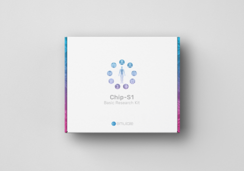
In this webinar, Lisa Chakrabarti, PhD, from Institut Pasteur described how her team developed a lymphoid Organ-Chip (LO Chip) using the Emulate Chip-S1® Stretchable Chip to address the challenge of predicting vaccine immunogenicity in humans. Dr. Chakrabarti went into detail about how the LO chip recreated key lymphoid tissue features, including T cell/B cell interactions and emigration of matured plasmablasts, and demonstrated the capability to assess responses to mRNA vaccines. 

Key highlights from this webinar include how: 

  • The model recreates lymphoid tissue features, including CD4+ T cell/B cell cluster formation and emigration of matured plasmablasts. 
  • The LO Chip effectively mimics vaccine boosts, amplifying SARS-CoV-2 Spike protein-specific B cells and antibody production.
  • The dynamically perfused culture system outperforms traditional 2D and 3D static cultures in immune response simulation. 
  • The LO Chip is responsive to both protein and mRNA-encoded antigens, highlighting its potential for preclinical evaluation of vaccine boosting strategies. 

Read Dr. Chakrabarti’s full paper here.

Modeling memory B cell responses in a lymphoid organ-chip to evaluate mRNA vaccine boosting

Organ Model: Lymphoid Follicle

Application: Vaccine development

How Organ-Chips Were Used: Predicting the immunogenicity of candidate vaccines in humans remains a challenge. To address this issue, the authors developed a Lymphoid Organ-Chip (LO Chip) based on a microfluidic chip seeded with human PBMCs at high density within a 3D collagen matrix. The LO Chip represents a versatile platform suited to the preclinical evaluation of vaccine boosting strategies.

Key highlights:

  • The model recreates key lymphoid tissue features, including CD4+ T cell/B cell cluster formation and emigration of matured plasmablasts.
  • It effectively mimics vaccine boosts, amplifying SARS-CoV-2 Spike protein-specific B cells, plasmablast differentiation and antibody production.
  • The dynamically perfused culture system outperforms traditional 2D and 3D static cultures in immune response simulation.
  • The LO Chip is responsive to both protein and mRNA-encoded antigens, highlighting its potential for preclinical evaluation of vaccine boosting strategies.

Products Used In This Publication

DNA origami vaccine (DoriVac) nanoparticles improve both humoral and cellular immune responses to infectious diseases

Organ Model: Lymph Node

Application: Immunology, Vaccine Development

Abstract: Current SARS-CoV-2 vaccines have demonstrated robust induction of neutralizing antibodies and CD4+ T cell activation, however CD8+ responses are variable, and the duration of immunity and protection against variants are limited. Here we repurposed our DNA origami vaccine platform, DoriVac, for targeting infectious viruses, namely SARS-CoV-2, HIV, and Ebola. The DNA origami nanoparticle, conjugated with infectious-disease-specific HR2 peptides, which act as highly conserved antigens, and CpG adjuvant at precise nanoscale spacing, induced neutralizing antibodies, Th1 CD4+ T cells, and CD8+ T cells in naïve mice, with significant improvement over a bolus control. Pre-clinical studies using lymph-node-on-a-chip systems validated that DoriVac, when conjugated with antigenic peptides or proteins, induced promising cellular immune responses in human cells. These results suggest that DoriVac holds potential as a versatile, modular vaccine platform, capable of inducing both humoral and cellular immunities. The programmability of this platform underscores its potential utility in addressing future pandemics.

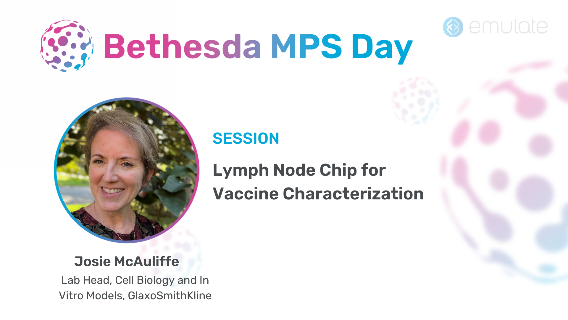
Lymph Node Chip for Vaccine Characterization

Featured session from Bethesda MPS Day, which took place on November 9, 2023.

Dr. Josie McAuliffe from GlaxoSmithKline (GSK) discusses her team’s efforts to employ a Lymph Node-Chip model to better understand and improve vaccine performance. Traditional vaccine development often relies on animal models and two-dimensional cell cultures that may not accurately predict clinical outcomes. The Lymph Node-Chip, a microphysiological system that recreates aspects of lymph node architecture and immune cell interactions, offers a more human-relevant environment for evaluating vaccine antigens and optimizing immune responses.

By collaborating with the Wyss Institute, Dr. McAuliffe’s team leveraged a Lymph Node-on-a-Chip platform capable of supporting T and B cells, as well as dendritic cells, under flow conditions. They demonstrated that this model could detect key immunological readouts, including cytokines/chemokines and antigen-specific antibody responses, after administration of a novel self-amplifying mRNA (SAM) vaccine formulation. The Lymph Node-Chip also showed follicle formation, an integral part of germinal center reactions critical for generating high-affinity antibodies.

Encouraged by these results, GSK decided to internalize the Lymph Node-Chip system. The goal is to evaluate proprietary vaccines more effectively in-house, reduce logistical complexities, and potentially correlate Lymph Node-Chip data with clinical results. Early in-house experiments have revealed promising markers, such as CXCL13, IP-10, and IL-15, known to be associated with effective vaccine-induced immune responses in humans. The team is now refining the model, looking into antibody detection methods, immunophenotyping, and single-cell sequencing to gain deeper insight into vaccine mechanisms of action and improve translation to clinical success.

Key learnings from this presentation include:

Enhanced translational relevance: The Lymph Node-Chip provides a more complex, physiological context than traditional 2D cultures or animal models, potentially improving predictions of human vaccine outcomes.

Robust immunological endpoints: The chip supports T and B cell interactions, follicle formation, and cytokine/chemokine production, enabling comprehensive analysis of germinal center reactions and antibody production.

Application to novel vaccines: By integrating SAM mRNA vaccines into the Lymph Node-Chip, the team observed antigen-specific antibody responses and immune markers reminiscent of those seen in human vaccine recipients.

In-house implementation: Bringing the Lymph Node-Chip technology internally allows GSK to test proprietary formulations, streamline logistics, and refine experimental parameters without external dependencies.

Future directions: Ongoing optimization focuses on strengthening endpoint assays (e.g., sensitive antibody detection), correlating Lymph Node-Chip data with clinical efficacy, and potentially exploring other lymphoid tissues or different species (like nonhuman primates) for enhanced translational insights.

Ectopic Lymphoid Follicle Formation and Human Seasonal Influenza Vaccination Responses Recapitulated in an Organ-on-a-Chip

Organ Model: Lymphoid Follicle

Application: Immunology

Abstract: Lymphoid follicles (LFs) are responsible for generation of adaptive immune responses in secondary lymphoid organs and form ectopically during chronic inflammation. A human model of ectopic LF formation will provide a tool to understand LF development and an alternative to non-human primates for preclinical evaluation of vaccines. Here, it is shown that primary human blood B- and T-lymphocytes autonomously assemble into ectopic LFs when cultured in a 3D extracellular matrix gel within one channel of a two-channel organ-on-a-chip microfluidic device. Superfusion via a parallel channel separated by a microporous membrane is required for LF formation and prevents lymphocyte autoactivation. These germinal center-like LFs contain B cells expressing Activation-Induced Cytidine Deaminase and exhibit plasma cell differentiation upon activation. To explore their utility for seasonal vaccine testing, autologous monocyte-derived dendritic cells are integrated into LF Chips. The human LF chips demonstrate improved antibody responses to split virion influenza vaccination compared to 2D cultures, which are enhanced by a squalene-in-water emulsion adjuvant, and this is accompanied by increases in LF size and number. When inoculated with commercial influenza vaccine, plasma cell formation and production of anti-hemagglutinin IgG are observed, as well as secretion of cytokines similar to vaccinated humans over clinically relevant timescales.