
Constructing a Complex Culture System of the Celiac Disease Intestinal Mucosal Barrier on Chip

Featured session at Netherlands MPS Day, which took place on November 15, 2023.

Joram Mooiweer from the University Medical Center Groningen presented his team’s work on modeling celiac disease using advanced human-relevant systems, including intestinal organoids and Organ-Chips. Celiac disease involves a complex interplay of genetic predisposition, immune responses (especially from tissue-resident T cells), and interactions with the intestinal epithelium and microbiota. By taking advantage of patient-derived tissue and induced pluripotent stem cells (iPSCs), the group aims to reconstruct these intricate processes in vitro.

Mooiweer highlights two approaches to modeling celiac disease. First, using patient biopsies, they isolate both intraepithelial lymphocytes (IELs) and intestinal stem cells. Culturing organoids derived from these stem cells together with IELs in a carefully optimized co-culture medium, they can recreate aspects of the cytotoxic activity associated with celiac disease—something that can be further manipulated using bispecific antibodies. These co-cultures maintain viability of both the epithelial and immune cells, enabling exploration of celiac-related immune-mediated epithelial damage.

Second, the group also uses iPSCs from patients and controls to generate small intestine-like epithelial tissues that self-organize into complex, 3D folded architectures when seeded on chip. With the help of growth factor gradients, they achieve a cell-type composition and maturation state that closely mirrors the human small intestine, including various specialized epithelial cells and mesenchymal cell types. This system is genetically tractable and can be further refined to incorporate inflammatory cues, immune cells, and eventually patient-specific microbiota under physiologically relevant conditions.

Ultimately, these multifaceted in vitro platforms will enable mechanistic insights into how genetic variants influence celiac disease onset and progression. They also provide a foundation for modeling interactions between epithelial cells, immune cells, and environmental triggers, paving the way for more predictive and personalized therapeutic strategies.

Key learnings from this presentation include:

  • Complex disease modeling: Celiac disease involves interactions among epithelial cells, immune cells (IELs), and environmental factors (gluten). Recreating these dynamics in vitro is essential to dissect underlying mechanisms.
  • Patient-derived systems: Using biopsies, organoids, and expanded IEL populations, the team can simulate immune-driven epithelial damage in controlled culture environments.
  • iPSC-derived intestinal tissue: iPSC-based models yield highly structured and diverse epithelial cell populations on a chip, reflecting small-intestine-like architecture and enabling genetic manipulation for studying disease-associated loci.
  • Synergistic modeling strategies: Combining adult-stem-cell-derived organoids, iPSC-based tissues, immune cells, and future microbiota co-cultures provides a comprehensive platform for understanding celiac pathophysiology.
  • Toward translational impact: These advanced human-relevant models aim to identify genetic factors and potential therapeutic targets, contributing to more tailored interventions in celiac disease and related intestinal disorders.

SCRIB controls apical contractility during epithelial differentiation

Organ Model: Intestine (Caco2)

Application: Model Development

Abstract: Although mutations in the SCRIB gene lead to multiple morphological organ defects in vertebrates, the molecular pathway linking SCRIB to organ shape anomalies remains elusive. Here, we study the impact of SCRIB-targeted gene mutations during the formation of the gut epithelium in an organ-on-chip model. We show that SCRIB KO gut-like epithelia are flatter with reduced exposed surface area. Cell differentiation on filters further shows that SCRIB plays a critical role in the control of apical cell shape, as well as in the basoapical polarization of myosin light chain localization and activity. Finally, we show that SCRIB serves as a molecular scaffold for SHROOM2/4 and ROCK1 and identify an evolutionary conserved SHROOM binding site in the SCRIB carboxy-terminal that is required for SCRIB function in the control of apical cell shape. Our results demonstrate that SCRIB plays a key role in epithelial morphogenesis by controlling the epithelial apical contractility during cell differentiation.

Improving In Vitro Cancer Modeling with Chip-A1

Webinar Abstract

Oncology drug candidates are currently the least likely type of therapeutic to succeed in clinical trials, with only 5.1% of Phase I candidates going on to receive FDA approval1. Understanding a tumor’s microenvironment is key to regulating cancer progression and developing more effective therapies—and Chip-A1 will give researchers this capability. 

In this August 15, 2023 webinar, Luke Dimasi, Senior Director of Product Management at Emulate, provided an overview of the Chip-A1 Accessible Chip, a new Organ-Chip consumable that expands the applications of Organ-on-a-Chip technology by allowing users to create thicker, multilayered tissues within the epithelial culture chamber and directly treat the tissue with topical or aerosolized drugs. Following this introduction, Elee Shimshoni, PhD, postdoctoral researcher at MIT, discussed how she and her former team from the Wyss Institute at Harvard used a prototype of Chip-A1 and found that it offered a new approach for studying epithelial-stromal interactions in Barrett’s Esophagus as well as the broader underlying mechanisms associated with esophageal cancer progression. 

During this webinar, the speakers discussed: 

  • New capabilities of Chip-A1 that expand the applications of Organ-on-a-Chip technology 
  • Why Chip-A1 improves in vitro modeling of epithelial-stromal interactions and tumor microenvironments 
  • A case study of using Chip-A1 to model Barrett’s Esophagus
  • How a Barrett’s Esophagus Chip-A1 model could potentially serve as a tool for personalized drug-response assessments between different patients or genetic subpopulations

About Organ-on-a-Chip Technology 

Organ-on-a-Chip technology is poised to deliver a paradigm shift in drug discovery. By emulating human physiology, Organ-Chips have the potential to increase the predictive power of preclinical modeling and advance more drugs to the clinic. Learn more about Organ-on-a-Chip technology by downloading our free eBook.

Improving Gene Therapy Development with Organ-on-a-Chip Technology

With the potential to bring cures to some of the world’s most devastating diseases, including cancer, muscular atrophy, and blindness, gene therapy is a cutting-edge area of research that is generating a tremendous amount of excitement. Download this free eBook to learn what gene therapy is, how it works, and why Organ-on-a-Chip technology could help gene therapy reach widespread use.

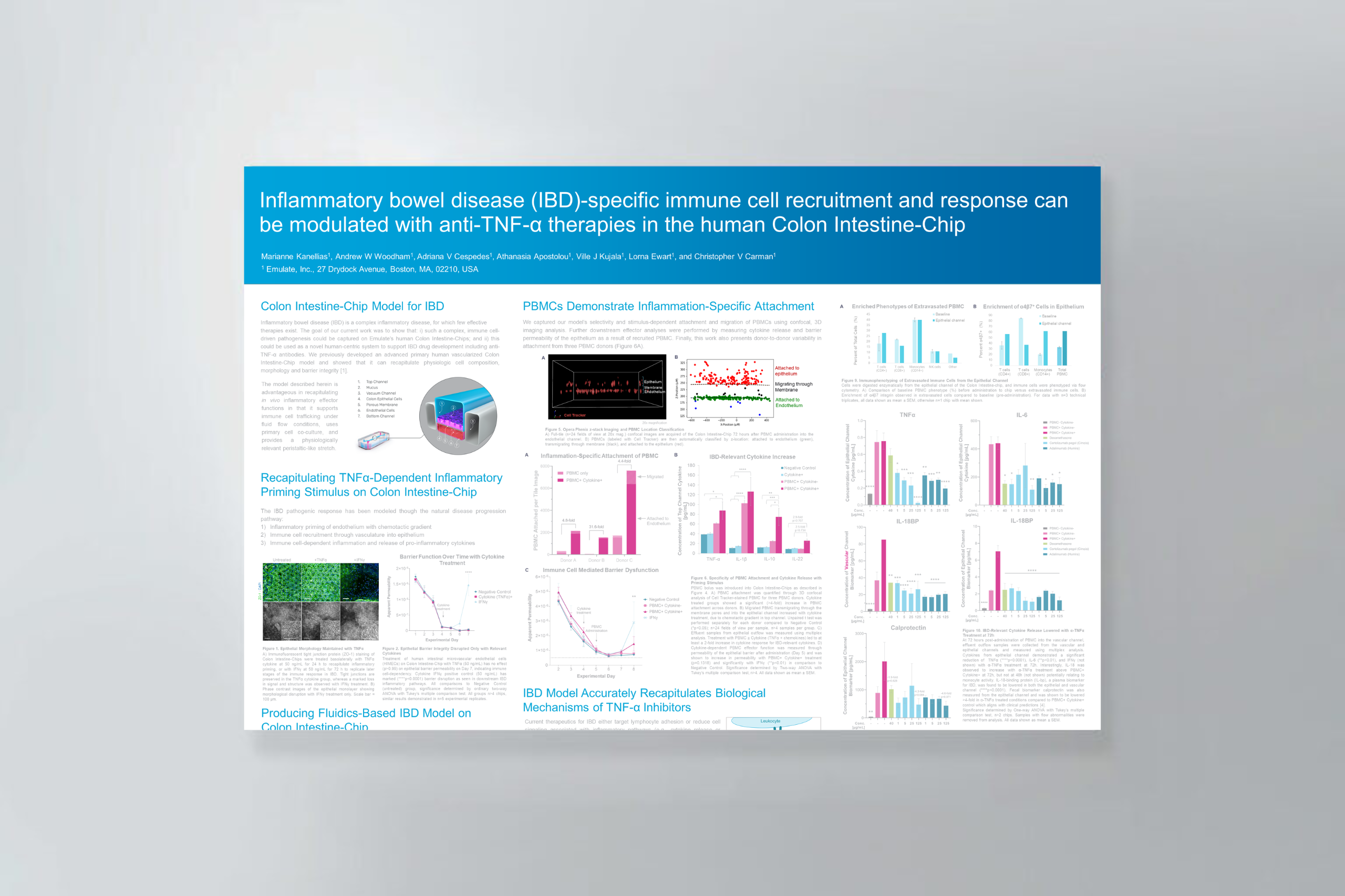
Inflammatory bowel disease (IBD)-specific immune cell recruitment and response can be modulated with anti-TNF-α therapies in the human Colon Intestine-Chip

Abstract

Inflammatory bowel disease (IBD) is a complex inflammatory disease, for which few effective therapies exist. The goal of our current work was to show that:

  1. Such a complex, immune cell-driven pathogenesis could be captured on Emulate’s human Colon Intestine-Chips
  2. This could be used as a novel human-centric system to support IBD drug development including anti-TNF-α antibodies. We previously developed an advanced primary human vascularized Colon Intestine-Chip model and showed that it can recapitulate physiologic cell composition, morphology and barrier integrity.

The model described in this poster is advantageous in recapitulating in vivo inflammatory effector functions in that it supports immune cell trafficking under fluid flow conditions, uses primary cell co-culture, and provides a physiologically relevant peristaltic-like stretch.

Microfluidic Model of Necrotizing Enterocolitis Incorporating Human Neonatal Intestinal Enteroids and a Dysbiotic Microbiome

Organ Model: Intestine (Necrotizing Enterocolitis)

Application: Immunology & Inflammation, Microbiome

Necrotizing enterocolitis (NEC) is a severe and potentially fatal intestinal disease that has been difficult to study due to its complex pathogenesis, which remains incompletely understood. The pathophysiology of NEC includes disruption of intestinal tight junctions, increased gut barrier permeability, epithelial cell death, microbial dysbiosis, and dysregulated inflammation. Traditional tools to study NEC include animal models, cell lines, and human or mouse intestinal organoids. While studies using those model systems have improved the field’s understanding of disease pathophysiology, their ability to recapitulate the complexity of human NEC is limited. An improved in vitro model of NEC using microfluidic technology, named NEC-on-a-chip, has now been developed. The NEC-on-a-chip model consists of a microfluidic device seeded with intestinal enteroids derived from a preterm neonate, co-cultured with human endothelial cells and the microbiome from an infant with severe NEC. This model is a valuable tool for mechanistic studies into the pathophysiology of NEC and a new resource for drug discovery testing for neonatal intestinal diseases. In this manuscript, a detailed description of the NEC-on-a-chip model will be provided.

Products Used In This Publication

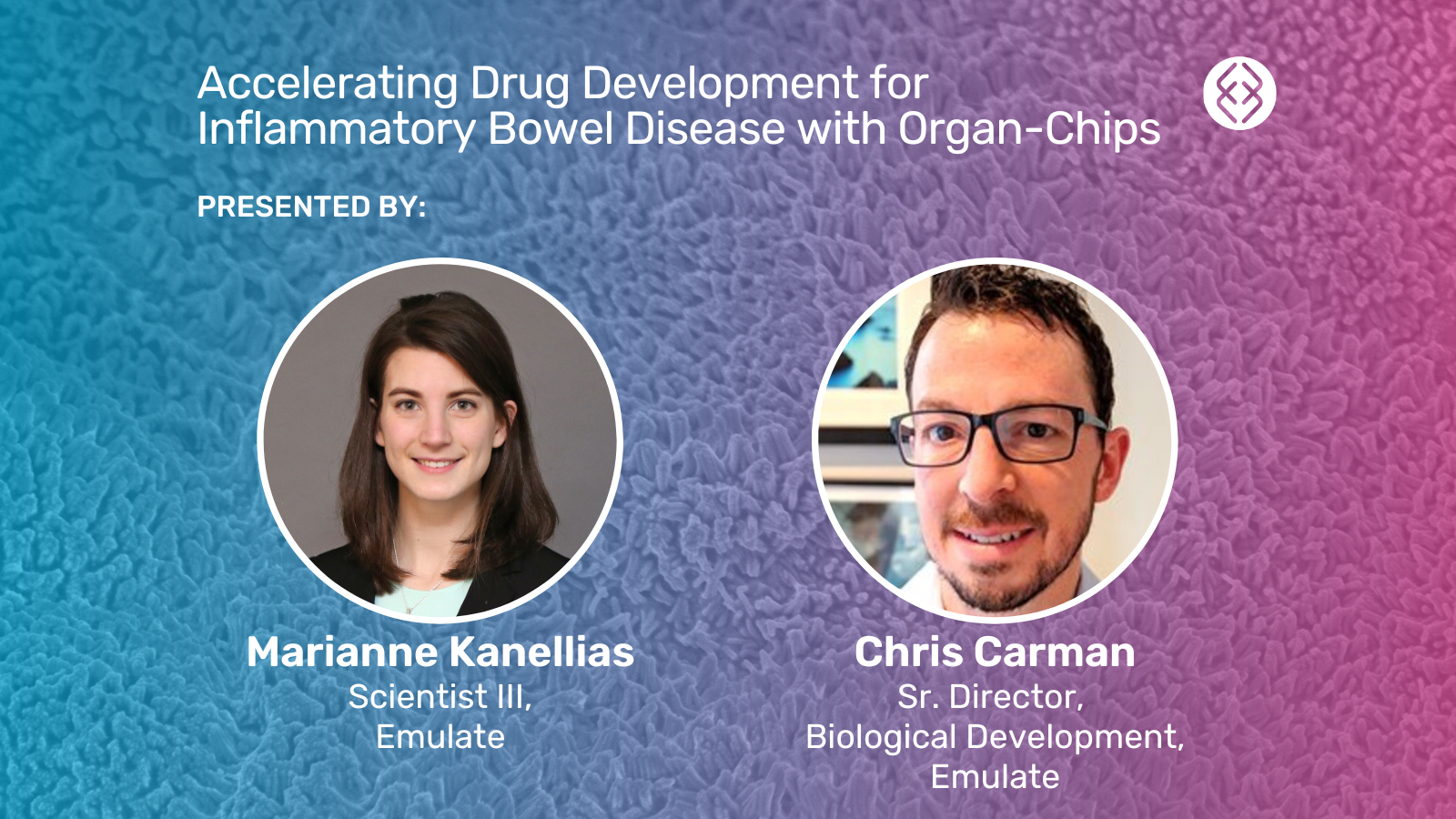
Accelerating Drug Development for Inflammatory Bowel Disease with Organ-Chips

Webinar Abstract

Inflammatory bowel disease (IBD) represents an enormous area of unmet medical need. Despite ~10 million people suffering from IBD globally, ~50% of patients fail to respond to on-market therapy, and ~90% fail to experience long-term remission.

The limited efficacy of IBD therapeutics is due in part to the limited human relevance of conventional preclinical models, which restrict researchers to studying just one aspect of IBD at a time. These models fail to accurately predict drug effects within the complex intestinal microenvironment, leading to high clinical trial failure rates.

It’s time for a human-centric approach to IBD drug discovery and development.

In this webinar, Marianne Kanellias discussed how researchers can use Emulate Organ-on-a-Chip technology to create a more representative and complex model of IBD pathogenesis. With the Emulate human Colon Intestine-Chip, researchers can recapitulate the inflammatory response of IBD—from immune cell vascular attachment, to migration, to downstream effector function and barrier damage—and evaluate the efficacy of anti-inflammatory therapeutics in a more human-relevant model of IBD.

Key highlights from this IBD webinar:

  • Common challenges in modeling inflammatory disease
  • Recreating gut- and inflammation-specific immune cell recruitment with the Colon Intestine-Chip
  • Evaluating clinically relevant IBD therapeutics across a range of mechanisms of action
  • A panel discussion and live Q&A with Marianne Kanellias and Chris Carman, PhD who led the development of the immune cell recruitment application for the Colon Intestine-Chip

For additional data, see the data in the inflammatory immune cell recruitment application note.

Dissecting Cancer-Microbiome Interactions with Organoids and Organ-Chips

Webinar Abstract

Over the past 15 years, organoids have revolutionized the study of human organ and tumor behavior. In the coming years, Organ-on-a-Chip technology promises to rapidly increase the complexity of human organotypic models, enabling the discovery of more physiologically relevant insights into human health and disease. 

In this on-demand webinar, Jens Puschhof, PhD, of the German Cancer Research Center (DKFZ) discusses how lessons learned from organoid biology can be applied to Organ-on-a-Chip research and examine areas where these two technologies are being combined with great potential for synergy. In particular, he shares aspects of his team’s work studying the impact of cancer-associated bacteria in colorectal cancer metastasis using Emulate Organ-Chip models. 

Key discussion points / learnings: 

  • The synergies that exist between organoids and Organ-on-a-Chip technology 
  • How insights from organoid research can be translated to Organ-Chips  
  • How Organ-Chips can be used to study the cancer-microbiome

Epithelial-Stromal Interactions in Barrett’s Esophagus Modeled in Human Organ Chips

Organ Model: Esophagus

Application: Cancer

Abstract: The pathogenesis of subsquamous intestinal metaplasia (SSIM), in which glands of Barrett’s esophagus (BE) are buried under esophageal squamous epithelium, is unknown. In a rat model of reflux esophagitis, we found that columnar-lined esophagus developed via a wound-healing process involving epithelial-mesenchymal plasticity (EMP) that buried glands under ulcerated squamous epithelium. To explore a role for reflux-induced EMP in BE, we established and characterized human Barrett’s organoids and sought evidence of EMP after treatment with acidic bile salts (AB). We optimized media to grow human BE organoids from immortalized human Barrett’s cells and from BE biopsies from seven patients, and we characterized histological, morphological, and molecular features of organoid development. Features and markers of EMP were explored following organoid exposure to AB, with and without a collagen I (COL1) matrix to simulate a wound-healing environment. All media successfully initiated organoid growth, but advanced DMEM/F12 (aDMEM) was best at sustaining organoid viability. Using aDMEM, organoids comprising nongoblet and goblet columnar cells that expressed gastric and intestinal cell markers were generated from BE biopsies of all seven patients. After AB treatment, early-stage Barrett’s organoids exhibited EMP with loss of membranous E-cadherin and increased protrusive cell migration, events significantly enhanced by COL1. Using human BE biopsies, we have established Barrett’s organoids that recapitulate key histological and molecular features of BE to serve as high-fidelity BE models. Our findings suggest that reflux can induce EMP in human BE, potentially enabling Barrett’s cells to migrate under adjacent squamous epithelium to form SSIM.NEW & NOTEWORTHY Using Barrett’s esophagus (BE) biopsies, we established organoids recapitulating key BE features. During early stages of organoid development, a GERD-like wound environment-induced features of epithelial-mesenchymal plasticity (EMP) in Barrett’s progenitor cells, suggesting that reflux-induced EMP can enable Barrett’s cells to migrate underneath squamous epithelium to form subsquamous intestinal metaplasia, a condition that may underlie Barrett’s cancers that escape detection by endoscopic surveillance, and recurrences of Barrett’s metaplasia following endoscopic eradication therapy.

An Unparalleled Window into IBD Immune Response with Organ-on-a-Chip Technology

Inflammatory bowel disease (IBD) is characterized by dysregulated immune response, ultimately leading to barrier damage. Unfortunately, conventional research models are not capable of capturing this complexity, resulting in an incomplete understanding of disease biology and high clinical trial attrition.

In this video, see how Emulate Organ-Chips offer an unparalleled window into IBD human immune response. By incorporating critical features of the human tissue microenvironment, Organ-Chips enable researchers to model the complexity of immune cell recruitment in a tissue-, disease-, and species-specific manner, and evaluate the efficacy of clinically relevant drug candidates.