With over 150 peer-reviewed publications across 30+ organ models, Emulate Organ-Chips are empowering researchers to make game-changing scientific breakthroughs! Download this digest to easily explore all publications related to your field of research, or to just learn more about how the technology itself is developing.
New this quarter:
Fallopian Tube
- Human fallopian tube-on-a-chip for preclinical testing of non-hormonal contraceptives with living human sperm
Intestine
- Probiotic intervention mitigates radiation-induced intestinal injury by alleviating oxidative stress in a human gut-on-a-chip
Lung (Airway)
- Development of an acute inhalation toxicity testing method based on a lung-on-a-chip
Lung (Alveolus)
- Enhanced lung delivery of an immunostimulatory duplex RNA augments the antitumor activity by reshaping systemic cytokine pharmacodynamics
Lung (Alveolus)
- Alveolus-on-a-Chip: A Novel Tool for Modeling Lung Transplant Cold Storage Ischemia/Reperfusion Injury
Placenta
- Simvastatin Restores Uteroplacental Hemodynamics and Trophoblast Function in Obstetric Antiphospholipid Syndrome in a Placenta-on-a-Chip Model
REVIEW ARTICLES:
Female Reproductive Tract
- Female Reproductive Tract Organ-on-Chips: Modeling Barrier Function and Drug Transport
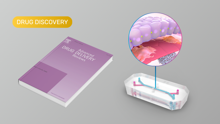
Drug Development
- Human organ-on-a-chip technology as a catalyst for drug discovery
PUBLISHED FROM PRE-PRINT:
Intestine (Colon)
- Human inflammatory bowel disease-on-a-chip for modelling disease progression, cancer initiation and sex-specific effects
Lymph Node
- In vitro recapitulation of intramuscular mRNA vaccination with naive and recall antigens using a human lymphoid follicle chip platform
Vasculature
- Human coronary artery organ-chip with circulating immune cells recapitulates anti-inflammatory effect of pulsatile wall strain