Synopsis
Lung transplantation is a life-saving treatment for patients with end-stage lung disease. Despite advances in transplantation technology, however, early graft injury remains a major challenge, underscoring the need for better insight into the biological processes that can compromise transplant success.
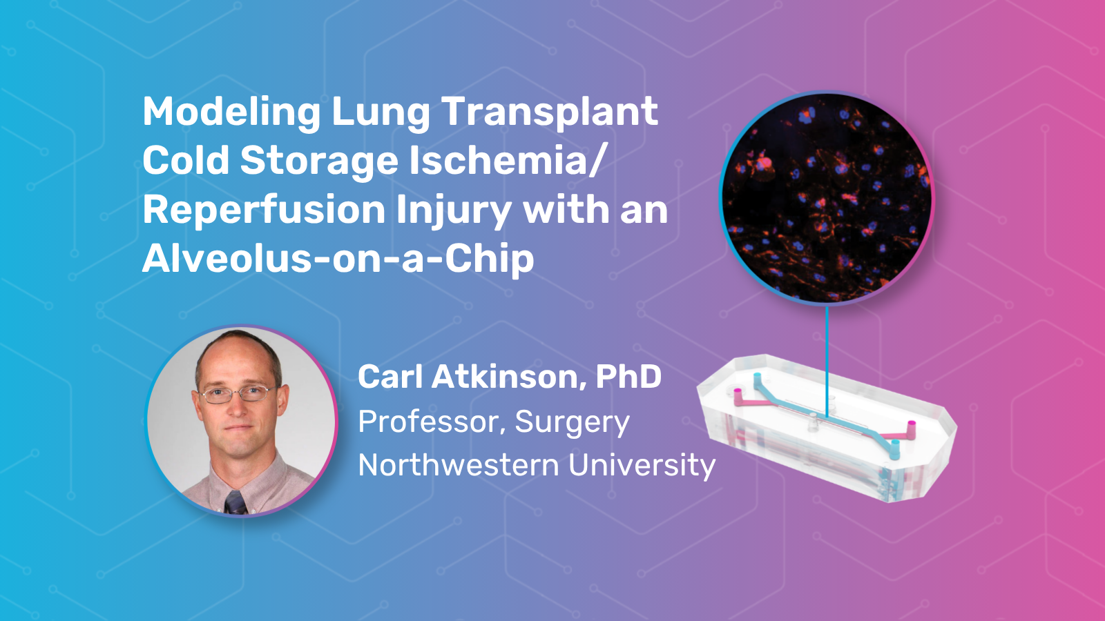
In this on-demand webinar, Carl Atkinson, PhD, Professor at Northwestern University, discusses his team’s recent publication describing a novel human Alveolus-on-a-Chip model of lung transplant cold storage ischemia/reperfusion injury. This type of injury can occur when donor lungs are preserved at low temperatures before transplant and then rewarmed and reperfused in the recipient, triggering inflammatory and barrier-disruptive responses that may contribute to poor early graft function.
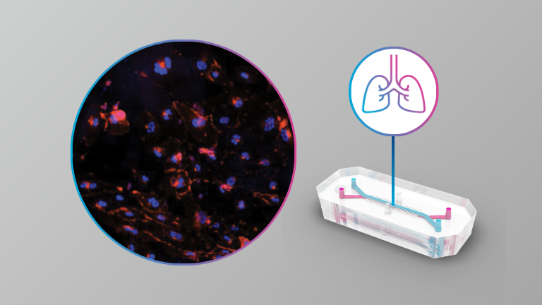
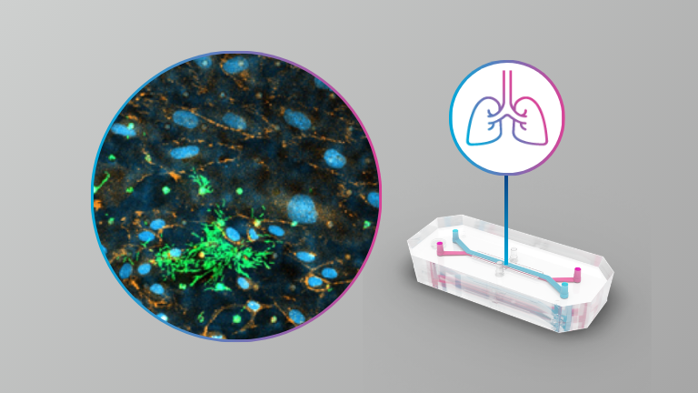
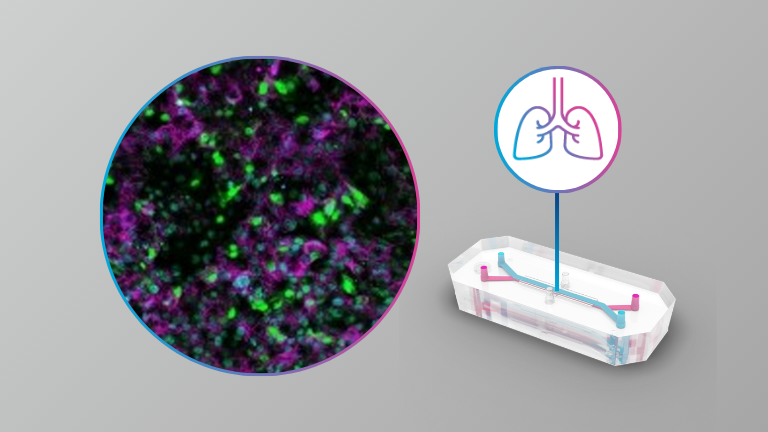
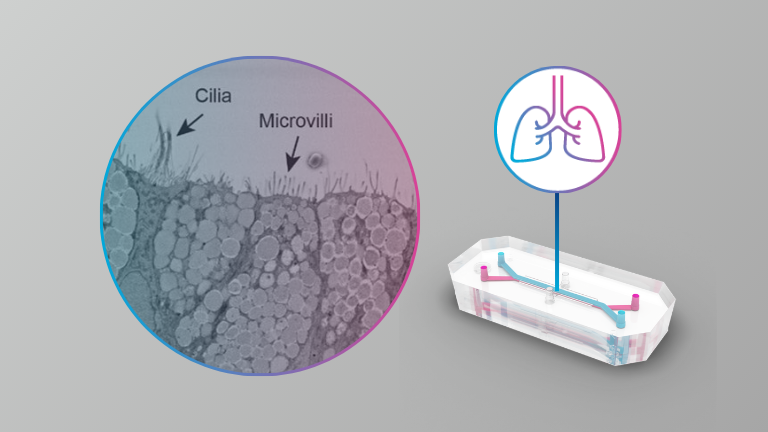
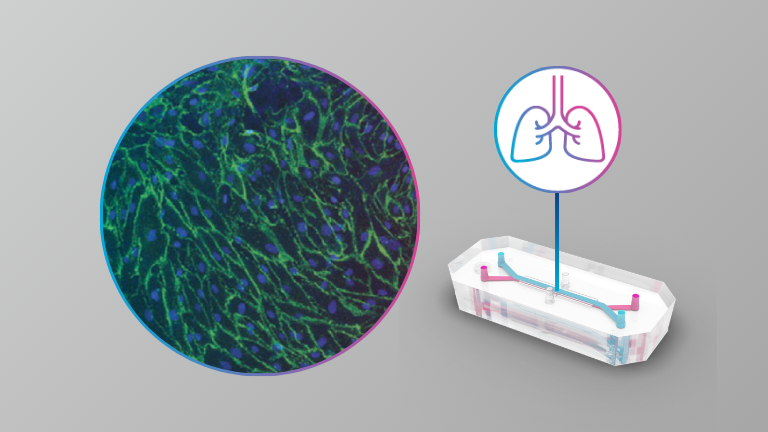
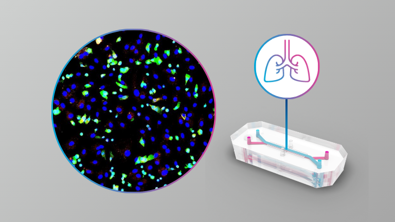
Using Emulate’s Organ-on-a-Chip technology, Dr. Atkinson’s team recreated key features of donor lung cold storage and reperfusion in a dynamic, human-relevant model of the alveolar-capillary interface. The model combines human alveolar epithelial cells and lung microvascular endothelial cells under an air-liquid interface, vascular-like flow, and cyclic mechanical stretch to better reflect the structure and function of the distal lung.
Dr. Atkinson shares how the model recapitulated clinically relevant hallmarks of lung transplant injury, including transient edema, disruption of epithelial and endothelial barrier markers, proinflammatory chemokine release, adhesion molecule regulation, and distinct epithelial and endothelial gene expression changes. The presentation highlights how the Alveolus-on-a-Chip model can help researchers investigate transplant-associated lung injury mechanisms and support future therapeutic discovery.
Key Learning Points
- What ischemia/reperfusion injury is and why it matters in the context of donor lung preservation and transplantation
- How a human Alveolus-on-a-Chip model can simulate lung transplant cold storage and reperfusion injury in vitro
- How cold storage ischemia/reperfusion injury impacts alveolar barrier function, endothelial integrity, and fluid accumulation in the chip
- How Organ-on-a-Chip models may accelerate mechanistic research and therapeutic screening in lung transplantation