Creating Bone-Chip Models to Discover New Insights into the Mechanisms of Breast Cancer Metastasis

Metastasis in breast cancer is a leading cause of mortality. In particular, breast cancer that metastasizes to the bone significantly worsens patient outcomes; however, the mechanisms underlying this process remain poorly understood. While previous studies have focused on how interactions between cancer cells and bone marrow contribute to cancer metastasis, significantly fewer studies have been completed that aim to understand how osteocytes—the primary regulators of the bone environment—influence metastatic breast cancer progression.

This case study summarizes how Dr. Stefaan Verbruggen and colleagues at Queen Mary University of London used Emulate Organ-Chips with the Human Emulation System to create a dynamic bone model of breast cancer metastasis, enabling them to elucidate the molecular mechanisms governing the interplay between cancer cells and osteocytes under conditions of mechanical loading.

In this case study, you will learn how:

  • Researchers used Organ-Chips to recapitulate breast cancer metastasis to the bone.
  • Mechanical stimulation plays a key role in early- and late-stage breast cancer metastasis.
  • Organ-Chips can benefit cancer researchers through enabling tissue co-cultures and the selective application of shear stress in a complex 3D tumor microenvironment.

A Novel Primary Cilium-Mediated Mechanism Through which Osteocytes Regulate Metastatic Behavior of Both Breast and Prostate Cancer Cells

Organ Model: Breast and prostate cancer bone metastasis model

Application: Cancer

Abstract: Bone metastases are a common cause of suffering in breast and prostate cancer patients, however, the interaction between bone cells and cancer cells is poorly understood. Using a series of co-culture, conditioned media, human cancer spheroid, and organ-on-a-chip experiments, this study reveals that osteocytes suppress cancer cell proliferation and increase migration via tumor necrosis factor alpha (TNF-α) secretion. This action is regulated by osteocyte primary cilia and associated intraflagellar transport protein 88 (IFT88). Furthermore, it shows that cancer cells block this mechanism by secreting transforming growth factor beta (TGF-β), which disrupts osteocyte cilia and IFT88 gene expression. This bi-directional crosstalk signaling between osteocytes and cancer cells is common to both breast and prostate cancer. This study also proposes that osteocyte inhibition of cancer cell proliferation decreases as cancer cells increase, producing more TGF-β. Hence, a positive feedback loop develops accelerating metastatic tumor growth. These findings demonstrate the importance of cancer cell-osteocyte signaling in regulating breast and prostate bone metastases and support the development of therapies targeting this pathway.

Bone Marrow Microenvironment-On-Chip for Culture of Functional Hematopoietic Stem Cells

Organ Model: Bone marrow (mouse)

Application: Model Development

Abstract: Hematopoiesis takes place in the bone marrow and is supported by a complex cellular and molecular network in the bone marrow microenvironment. Commonly used models of the human bone marrow microenvironment include murine models and two-dimensional and three-dimensional tissue cultures. While these model systems have led to critical advances in the field, they fail to recapitulate many aspects of the human bone marrow. This has limited our understanding of human bone marrow pathophysiology and has led to deficiencies in therapy for many bone marrow pathologies such as bone marrow failure syndromes and leukemias. Therefore, we have developed a modular murine bone marrow microenvironment-on-chip using a commercially available microfluidic platform. This model includes a vascular channel separated from the bone marrow channel by a semi-porous membrane and incorporates critical components of the bone marrow microenvironment, including osteoblasts, endothelial cells, mesenchymal stem cells, and hematopoietic stem and progenitor cells. This system is capable of maintaining functional hematopoietic stem cells in vitro for at least 14 days at frequencies similar to what is found in the primary bone marrow. The modular nature of this system and its accessibility will allow for acceleration of our understanding of the bone marrow.

Mechanical Stimulation Modulates Osteocyte Regulation of Cancer Cell Phenotype

Organ Model: Breast and prostate cancer bone metastasis model

Application: Cancer

Abstract: Breast and prostate cancers preferentially metastasise to bone tissue, with metastatic lesions forming in the skeletons of most patients. On arriving in bone tissue, disseminated tumour cells enter a mechanical microenvironment that is substantially different to that of the primary tumour and is largely regulated by bone cells. Osteocytes, the most ubiquitous bone cell type, orchestrate healthy bone remodelling in response to physical exercise. However, the effects of mechanical loading of osteocytes on cancer cell behaviour is still poorly understood. The aim of this study was to characterise the effects of osteocyte mechanical stimulation on the behaviour of breast and prostate cancer cells. To replicate an osteocyte-controlled environment, this study treated breast (MDA-MB-231 and MCF-7) and prostate (PC-3 and LNCaP) cancer cell lines with conditioned media from MLO-Y4 osteocyte-like cells exposed to mechanical stimulation in the form of fluid shear stress. We found that osteocyte paracrine signalling acted to inhibit metastatic breast and prostate tumour growth, characterised by reduced proliferation and invasion and increased migration. In breast cancer cells, these effects were largely reversed by mechanical stimulation of osteocytes. In contrast, conditioned media from mechanically stimulated osteocytes had no effect on prostate cancer cells. To further investigate these interactions, we developed a microfluidic organ-chip model using the Emulate platform. This new organ-chip model enabled analysis of cancer cell migration, proliferation and invasion in the presence of mechanical stimulation of osteocytes by fluid shear stress, resulting in increased invasion of breast and prostate cancer cells. These findings demonstrate the importance of osteocytes and mechanical loading in regulating cancer cell behaviour and the need to incorporate these factors into predictive in vitro models of bone metastasis.

Bone Chip System to Monitor Osteogenic Differentiation Using Optical Imaging

Published in: Microfluidics and Nanofluidics

Abstract

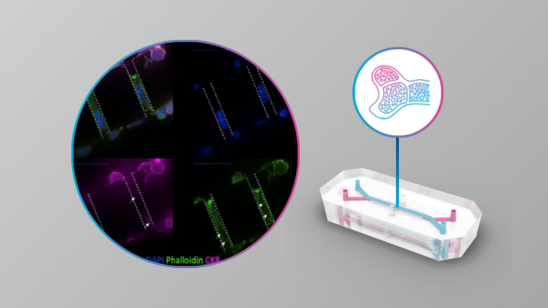
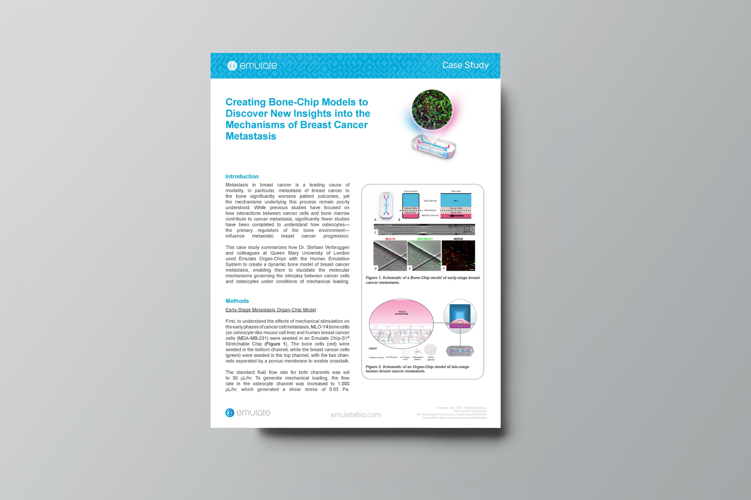
Human organoids and Organ-on-Chip systems to predict human responses to new therapies and for the understanding of disease mechanisms are being more commonly used in translational research. We have developed a bone-chip system to study osteogenic differentiation in vitro, coupled with optical imaging approach which provides the opportunity of monitoring cell survival, proliferation and differentiation in vitro without the need to terminate the culture. We used the mesenchymal stem cell (MSC) line over-expressing bone morphogenetic protein-2 (BMP-2), under Tet-Off system, and luciferase reporter gene under constitutive promoter. Cells were seeded on chips and supplemented with osteogenic medium. Flow of media was started 24 h later, while static cultures were performed using media reservoirs. Cells grown on the bone-chips under constant flow of media showed enhanced survival/proliferation, comparing to the cells grown in static conditions; luciferase reporter gene expression and activity, reflecting the cell survival and proliferation, was quantified using bioluminescence imaging and a significant advantage to the flow system was observed. In addition, the flow had positive effect on osteogenic differentiation, when compared with static cultures. Quantitative fluorescent imaging, performed using the osteogenic extra-cellular matrix-targeted probes, showed higher osteogenic differentiation of the cells under the flow conditions. Gene expression analysis of osteogenic markers confirmed the osteogenic differentiation of the MSC-BMP2 cells. Immunofluorescent staining performed against the Osteocalcin, Col1, and BSP markers illustrated robust osteogenic differentiation in the flow culture and lessened differentiation in the static culture. To sum, the bone-chip allows monitoring cell survival, proliferation, and osteogenic differentiation using optical imaging.