Chip Cradle Protocol (Supplement to Organ-Chip Culture Protocols)

Introduction

This is a supplemental protocol which outlines the use of the new Chip Cradle. It is not intended to replace current Organ-Chip protocols, but to demonstrate how the new cradle integrates into existing Organ-Chip protocols.

This user guide contains important information to safely and effectively use the Chip Cradle. All users should thoroughly read and understand this guide before use.

Fixed Chip Imaging Adapter User Guide

Introduction

The Fixed Chip Imaging Adapter organizes fixed chips for high-throughput imaging and ensures compatibility with SBS footprint advanced imaging equipment. It can also prevent evaporation for 12+ hours when used with off-the-shelf accessories.

This user guide details what is included with the kit, as well as different ways to configure the adapter to prevent evaporation for different periods.

Compound Distribution Kit Protocol

Introduction

In both in vitro and in vivo experiments, researchers must consider compound distribution within the biological model and experimental setup prior to quantitative drug studies, as distribution determines exposure — the concentration of a compound that cells truly experience.

In in vivo systems, this is addressed by volume of distribution studies, which relate compound dosage to its effective concentration. However, in both in vivo and in vitro studies, the distribution effects of system components, such as infusion tubing, syringes, tissue-culture plates, and pipette tips, are often missed.

With Organ-Chip experiments, we proactively address compound distribution in a number of ways. Several of these are embedded in our protocol designs, where we have selected experimental conditions to optimize compound exposure. Additionally, we have developed the Compound Distribution Kit to directly evaluate distribution and compound exposure.

The Compound Distribution Kit is intended to be used as a specialized control experiment — the distribution control experiment — prior to the intended Organ-Chip study. As such, the contents of the Compound Distribution Kit mirror the contents of the Organ-Chip Bio-Kit, and the protocol used for the distribution control experiment mirrors a simplified version of the intended study (e.g., without cells or ECM coating). The distribution control experiment’s output indicates whether any compound may be distributed into the system and away from cells. Moreover, in some cases, the distribution control can be used to quantitatively correct the experimental results of the intended study and assign it appropriate error bars.

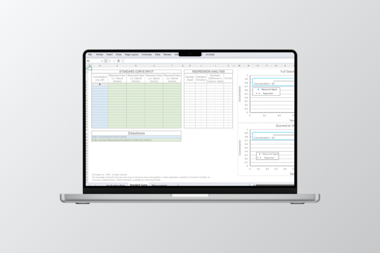
Standard Curve Calculator

Version 1.0 (Release Date: November 2021)​

Introduction

The Standard Curve Calculator is an Excel-based solution used for calculating a standard curve from raw signal measurements, such as optical density or fluorescence intensity from a plate reader assay. The conversion of raw signal intensity to a concentration (or other known readout) is performed using a standard curve. A typical use case is calculating the concentration of a given test molecule in Organ-Chip effluent. However, this calculator applies broadly to any raw readout or signal. ​

Glucose Quantification Assay

Introduction

This assay provides a simple and sensitive method to quantify glucose levels from Organ-Chip effluent samples.

Barrier Function Readout Analysis

Introduction

The maintenance or disruption of tissue barriers is an essential part of the pathophysiology of many diseases. The ability to quantitatively characterize tissue barrier is critical in the evaluation of barrier integrity and function.

This protocol is to be used to assess the permeability of an Organ-Chip’s endothelial-epithelial barrier. Apparent permeability (Papp) of tracer molecules is determined by dosing the inlet of one channel, collecting the effluent of both channels, and calculating the amount of compound that crossed through the membrane over time. See full method below and associated Papp Calculator (EC004) for data analysis.

Live Staining of CLF Uptake into Bile Canaliculi

Introduction

Corning® Cholyl-lysyl-fluorescein (CLF) staining is used to visualize the structure and function of bile canaliculi in polarized hepatocytes. CLF is a substrate for the canalicular bile salt export pump (BSEP), thus it can be used to visualize BSEP-mediated canalicular efflux, as well as to label the bile canaliculi structures.

Gsh-Glo™ Glutathione Assay

Introduction

The GSH-Glo™ Assay is a luminescent-based assay for the detection and quantification of glutathione (GSH), an antioxidant that can prevent damage to cellular components caused by reactive oxygen species such as free radicals, peroxides, and lipid peroxides. GSH is involved in the detoxification of both xenobiotic and endogenous compounds. A change in GSH levels can be used as an indicator of toxicity.

Glycogen Quantification Assay

Introduction

This protocol covers quantification of glycogen on our Organ-Chips.

Triglycerides Quantification Assay