Abstract: Conventional dogma suggests that decompression sickness (DCS) is caused by nitrogen bubble nucleation in the blood vessels and/or tissues; however, the abundance of bubbles does not correlate with DCS severity. Since immune cells respond to chemical and environmental cues, we hypothesized that the elevated partial pressures of dissolved gases drive aberrant immune cell phenotypes in the alveolar vasculature. To test this hypothesis, we measured immune responses within human lung-on-a-chip devices established with primary alveolar cells and microvascular cells. Devices were pressurized to 1.0 or 3.5 atm and surrounded by normal alveolar air or oxygen-reduced air. Phenotyping of neutrophils, monocytes, and dendritic cells as well as multiplexed ELISA revealed that immune responses occur within 1 h and that normal alveolar air (i.e., hyperbaric oxygen and nitrogen) confer greater immune activation. This work strongly suggests innate immune cell reactions initiated at elevated partial pressures contribute to the etiology of DCS.
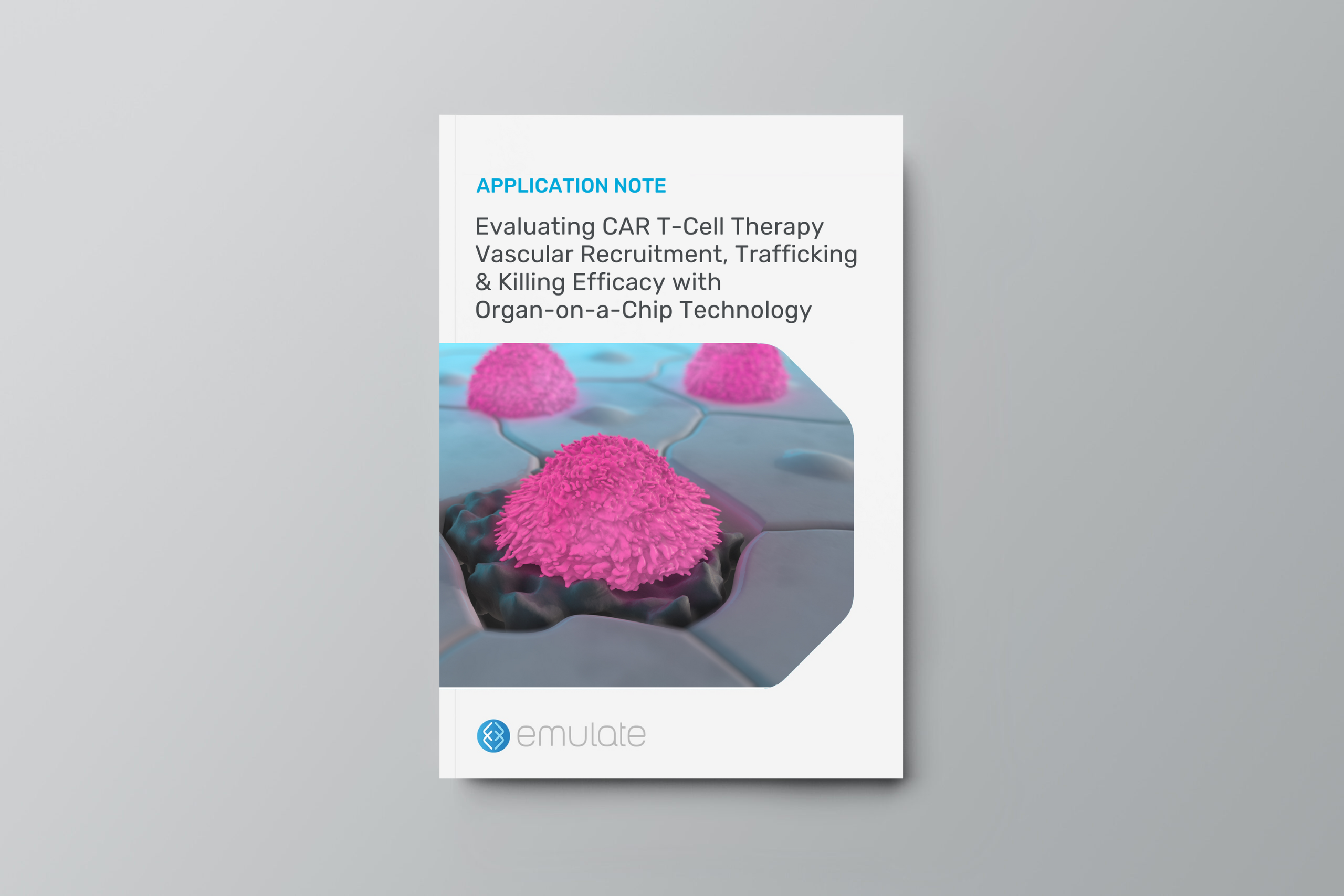
Chimeric antigen receptor (CAR) T-cell therapy shows significant potential in treating various human cancers. Unfortunately, researchers have faced difficulty in adapting this therapy to target solid tumors, where CAR T cells face unique challenges, such as antigen heterogeneity, an immuno-suppressive microenvironment, and T-cell exhaustion.
However, none of these challenges matter unless the CAR T cells can extravasate out of the blood stream and successfully migrate to the site of the tumor—an often-overlooked yet critical rate-limiting part of the process that cannot be modeled in conventional cell cultures.
This application note explains how Organ-Chips can be used to model the entire journey of solid tumor CAR T-cell therapy in a vascularized cancer cell line model.
In this application note, you will learn how Organ-Chips can be used to:
Evaluate CAR T-cell vascular recruitment and killing efficacy in a single, unified assay.
Assess antigen-specific killing and degranulation through a range of imaging and effluent-based analysis.
Investigate co-therapeutic efficacy, as shown by proof-of-concept IL-2 data.
Adapt the workflow to study a diverse range of solid tumor cell lines, immunotherapies, and co-therapeutics.
In this session, our expert speakers highlight how they have used Organ-Chips to study viral and bacterial infection, including SARS-CoV-2, Nipah virus, and Mycobacterium tuberculosis. Watch to learn how you can gain deeper insights into infectious disease pathogenesis, infection-induced inflammatory response, and more.
Talk 1: Human Lung Microfluidic Chip: Nipah Virus Disease Modeling and Antiviral Treatments in Maximum Containment
Sushma Bhosle, PhD
Molecular Virology Associate Study Director
NIH/NIAID
Dr. Bhosle’s research group at the National Institute of Allergy and Infectious Diseases (NIAID) leveraged human Small Airway Lung-Chips to model Nipah (NiV) virus disease modelling in maximum containment. They demonstrated the application of NiV-infected Small Airway Lung-Chips towards therapeutic evaluation of antiviral drugs. Further, successful recapitulation of neutrophil infiltration, critical immune responses, activation of endothelium leading to inflammation was achieved with human Lung-Chips.
Talk 2: Early events in tuberculosis—harnessing microphysiological models to study humanity’s oldest foe
Vivek Thacker, PhD
Group Leader
University of Heidelberg Medical Facility
Tissue microenvironments profoundly influence infection and treatment outcomes; but their roles can be difficult to dissect in a mechanistic manner. This is particularly so for tuberculosis, whose early stages of infection in alveoli are difficult to study in any animal model and not recapitulated in typical in vitro models of infection. In this talk, Dr. Thacker will describe how microphysiological models such as Organ-Chips are powerful tools to fill this gap and provide new insights into how obligate pathogens, such as Mycobacterium tuberculosis, adapt to specific tissue niches and what consequences this may have for treatment outcomes.
For this research, Dr. Thacker and team received the 2024 SwissTB Award.
Talk 3: Application of Airway-on-Chip Models to study Bacterial Lung Infection
Amy Ryan, PhD
Associate Professor of Anatomy and Cell Biology
University of Iowa
Many lung diseases, both acute and chronic, are associated with bacterial infection damaging the integrity of the epithelial barrier. Dr. Ryan’s recent collaborative research has developed Airway-on-chip models, which offer a microscale platform that mimics many features of human lung physiology. This platform facilitates the investigation of bacterial lung infections by replicating key features of the airway environment, including directional air flow and stretch. These models enable real-time monitoring of bacterial behavior and host responses, advancing our understanding of infection dynamics and the development of targeted therapeutic strategies.
Despite decades of therapeutic progress, respiratory viruses continue to pose a significant threat to global health, causing millions of deaths each year. A major challenge has been the limitations of conventional research models. Static2D cell culture and animal models often fall short in replicating disease pathology and therapeutic effect as they would appear in humans, hindering the development of infectious disease therapeutics and, in turn, leading to a continued health burden.
Fortunately, Organ-Chips can provide more human-relevant insights into infectious disease pathology and therapeutic efficacy. This case study explores three peer-reviewed studies in which researchers from the Wyss Institute at Harvard University used Organ-Chips to:
Rapidly identify promising treatments
Investigate how viruses evolve through human-to-human transmission
Study the role of mechanical forces on the innate immune response to viral infection
Abstract: The COVID-19 pandemic has highlighted the importance of physiologically relevant in vitro models to assist preclinical research. Here, we describe the adaptation of a human alveolus microphysiological system (MPS) model consisting of primary human alveolar epithelial and lung microvascular endothelial cells to study infection with SARS-CoV-2 at Biosafety Level 3 (BSL3) facility. This infection model recapitulates breathing-like stretch and culture of epithelial cells at the air-liquid interface (ALI) and resulted in clinically relevant cytopathic effects including cell rounding of alveolar type 2 cells (AT2) and disruption of the tight junction protein occludin (OCLN). Viral replication was confirmed by immunocytochemical nucleocapsid staining in the epithelium and increased shedding of SARS-CoV-2 virus within two days post-infection, associated with changes in innate host immune responses. Together, these data demonstrate that, under the experimental conditions used in this work, this human alveolus MPS chip can successfully model SARS-CoV-2 infection of human alveolar lung cells.
Featured session at Netherlands MPS Day, which took place on 11/15/2023.
Dr. Anne van der Does from Leiden University Medical Center presents work on modeling both airway and alveolar regions of the human lung using advanced in vitro systems, including Organ-on-a-Chip technology. The lung, a complex and heterogeneous organ, is composed of distinct epithelial cell populations and is continuously subjected to mechanical forces such as airflow and stretching during breathing. These factors differ between the large airways, small airways, and alveoli, and they strongly influence cellular maturation, function, and repair.
Her team’s research demonstrates that incorporating mechanical cues (e.g., airflow and cyclic stretch) into an airway epithelial chip model accelerates mucociliary clearance and enhances the directional beating of cilia, achieving a more physiologically mature state within a shorter timeframe than conventional static cultures. Similarly, alveolar region models are being developed using primary alveolar type II cells expanded in organoids and induced pluripotent stem cell (iPSC)-derived alveolar-like cells. This approach enables large-scale, long-term studies and even genetic manipulation via CRISPR-based gene editing. The goal is to create improved disease models for conditions like chronic obstructive pulmonary disease (COPD) and pulmonary fibrosis, incorporating additional cell types (endothelial and stromal cells), mechanical factors, and environmental exposures (e.g., cigarette smoke). Ultimately, these refined, human-relevant models allow more accurate investigation into lung development, disease progression, and the efficacy of potential therapeutics.
Key learnings from this presentation include:
Physiological relevance of mechanical cues: Integrating airflow and stretching significantly improves airway epithelial maturation, enhancing mucociliary clearance and cilia orientation within a shorter culture period compared to static conditions.
Complex lung modeling: Distinct lung regions (airways vs. alveoli) require unique culturing strategies. Region-specific cell isolation, differentiation protocols, and careful choice of mechanical parameters are essential for robust, functional models.
Biological insights into lung diseases: Improved in vitro models, including those derived from primary or iPSC-derived alveolar cells, facilitate the study of chronic lung diseases and repair mechanisms at the cellular and tissue level.
Scalability and genetic manipulation: Expanding primary alveolar cells and using iPSCs allow for large-scale disease modeling and gene editing approaches, enabling systematic exploration of disease-relevant genetic variants and testing of therapeutic strategies.
Towards multi-cellular, more complex systems: Incorporating additional lung cell types (such as endothelial cells and stromal cells) and modeling pathological conditions (fibrosis, alveolar damage) paves the way for more comprehensive and predictive human lung models.
Featured session at Bethesda MPS Day, which took place on November 9, 2023.
Dr. Sushma Bhosle from the Integrated Research Facility (IRF) at NIAID/NIH presents her work on using Lung-Chip models in maximum biosafety containment (BSL-4) settings for studying high-risk viruses such as Nipah virus (NiV). Traditional preclinical models (e.g., simple 2D cell cultures or animal models) often fail to accurately predict human clinical outcomes, and there’s a need for more physiologically relevant, human-based systems. Microphysiological systems (MPS), like the Emulate Small Airway Lung-Chip, provide a complex, human-relevant platform that incorporates airway epithelium and microvascular endothelium under air-liquid interface and flow conditions.
Dr. Bhosle’s team validated a Small Airway Lung-Chip using human primary cells, confirming proper differentiation and expression of markers indicative of mature airway tissue. They infected these chips with Nipah virus under BSL-4 conditions and monitored viral replication, barrier integrity, and immune responses. Compared to traditional transwell assays, the Airway Lung-Chip supported robust viral replication across both apical and basal compartments, displayed increased barrier permeability changes, and allowed detailed observation of cytokine profiles and endothelial inflammation. Treatment with antiviral drugs, such as zotatifin (an mRNA translation inhibitor) and remdesivir, reduced viral burden and restored barrier function. The chips also enabled the study of donor-to-donor variability in immune and inflammatory responses, reflecting the personalized aspects of human disease.
Furthermore, incorporation of immune cells (neutrophils) into the chip environment showed that immune cell recruitment and cytokine release patterns differed markedly upon viral infection, emphasizing the importance of including immune components for a more comprehensive disease model. This approach sets the stage for using Lung-Chip systems in maximum containment labs to test therapeutics, explore patient-specific responses, and understand high-risk virus biology with greater predictive value for clinical outcomes.
Key learnings from this presentation include:
High containment feasibility: The Airway Lung-Chip is successfully operated in BSL-4 conditions, enabling the study of severe pathogens like Nipah virus.
Physiological complexity: Compared to transwells, MPS offer dynamic air-liquid interface, fluid flow, and mechanical stretch, better replicating human lung physiology and yielding more meaningful data on viral infection and tissue responses.
Antiviral interventions: Testing antivirals directly on the chip shows how candidate drugs (zotatifin, remdesivir) impact viral replication, barrier integrity, and cytokine profiles in a human-relevant context.
Donor variability and immune involvement: The system captures variations in immune and inflammatory markers between donors and highlights the critical role of immune cell infiltration in shaping virus-induced pathology.
Broad potential applications: Beyond Nipah virus, these methods can extend to studying various high-risk pathogens, informing therapeutic development, and advancing precision medicine approaches in infectious diseases.
Featured session at Bethesda MPS Day, which took place on November 9, 2023.
Dr. Dylan Fudge from the U.S. Army DEVCOM Chemical Biological Center discusses the use of Organ-Chip models, specifically Emulate’s MPS platforms, to advance the Army’s predictive toxicology efforts related to chemical and biological threats. Traditional safety and efficacy assessments often rely heavily on animal models and simplistic in vitro assays, which may not accurately reflect human physiology, particularly for acute, high-risk agents. By integrating human-relevant cells into microfluidic devices, Dr. Fudge’s team aims to improve mechanistic understanding, dose-response assessments, and long-term impact evaluations of nerve agents, PFAS compounds, and pathogens like SARS-CoV-2.
He showcases three major projects:
VX Nerve Agent on Liver-Chip: Exposing a human Liver-Chip (with hepatocytes, endothelial cells, stellates, and Kupffer cells) to sub-lethal VX concentrations allowed for multi-omics analyses (proteomics, metabolomics, and transcriptomics). The system revealed shifts in metabolic pathways, bioenergetics, and nitrogen metabolism consistent with in vivo and clinical literature. Importantly, VX’s known mechanism—disruption of acetylcholine breakdown—was mirrored in altered choline metabolism, reinforcing the Liver-Chip’s translational relevance.
PFAS Exposure in Kidney and Liver-Chips: Investigating polyfluoroalkyl substances (PFAS), the team saw patterns of compound retention and toxicity aligned with known clinical effects. Liver and Kidney-Chips demonstrated differential absorption and metabolic responses to PFAS, supported by robust omics data. Oxygen stress markers and biomarkers like uric acid levels recapitulated known toxicity profiles, indicating the chips’ capacity to model chronic, low-level exposures and related subtler health effects.
SARS-CoV-2 Infection Using Lung-Chips: Lung-Chips incorporating airway and alveolar regions enabled controlled infection with SARS-CoV-2 variants. Despite minimal acute cytotoxicity, omics analyses detected hallmark inflammatory pathways and mechanistic insights matching emerging clinical findings—such as immune activation and potential neuro-related disease pathways—emphasizing the chip’s ability to reveal complex host-pathogen interactions.
Collectively, these studies highlight the versatility and reliability of Emulate’s Organ-Chip platforms under rigorous defense-oriented research conditions. Their capacity to maintain viable tissues, capture human-like biology, and provide high-content data (via omics) positions these systems as valuable tools to reduce animal use, support therapeutic development, and enhance understanding of chemical and biological threat agents at a mechanistic level.
Abstract: Mycobacterium tuberculosis (Mtb) cultured axenically without detergent forms biofilm-like cords, a clinical identifier of virulence. In lung-on-chip (LoC) and mouse models, cords in alveolar cells contribute to suppression of innate immune signaling via nuclear compression. Thereafter, extracellular cords cause contact-dependent phagocyte death but grow intercellularly between epithelial cells. The absence of these mechanopathological mechanisms explains the greater proportion of alveolar lesions with increased immune infiltration and dissemination defects in cording-deficient Mtb infections. Compression of Mtb lipid monolayers induces a phase transition that enables mechanical energy storage. Agent-based simulations demonstrate that the increased energy storage capacity is sufficient for the formation of cords that maintain structural integrity despite mechanical perturbation. Bacteria in cords remain translationally active despite antibiotic exposure and regrow rapidly upon cessation of treatment. This study provides a conceptual framework for the biophysics and function in tuberculosis infection and therapy of cord architectures independent of mechanisms ascribed to single bacteria.
Abstract: Acute exposure to high-dose gamma radiation due to radiological disasters or cancer radiotherapy can result in radiation-induced lung injury (RILI), characterized by acute pneumonitis and subsequent lung fibrosis. A microfluidic organ-on-a-chip lined by human lung alveolar epithelium interfaced with pulmonary endothelium (Lung Alveolus Chip) is used to model acute RILI in vitro. Both lung epithelium and endothelium exhibit DNA damage, cellular hypertrophy, upregulation of inflammatory cytokines, and loss of barrier function within 6 h of radiation exposure, although greater damage is observed in the endothelium. The radiation dose sensitivity observed on-chip is more like the human lung than animal preclinical models. The Alveolus Chip is also used to evaluate the potential ability of two drugs – lovastatin and prednisolone – to suppress the effects of acute RILI. These data demonstrate that the Lung Alveolus Chip provides a human relevant alternative for studying the molecular basis of acute RILI and may be useful for evaluation of new radiation countermeasure therapeutics