Synopsis
Organ-on-a-Chip technology is emerging as a powerful New Approach Methodology (NAM) for drug development, driven by the need for more human-relevant and scalable experimental models. As these systems move toward broader adoption, a key challenge remains: generating consistent, interpretable data that supports confident experimental and translational decision-making.
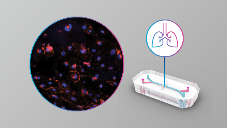
This webinar examines how imaging and AI-driven analysis workflows enable Organ-Chip studies to scale from innovation to routine application. Speakers begin with an overview of Organ-on-a-Chip technology and its role in addressing translational gaps in drug discovery, highlighting how Liver-Chips are being evaluated in collaboration with regulatory agencies for better prediction of drug-induced liver injury.
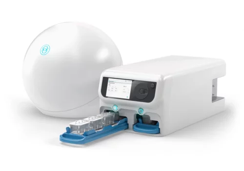
The session then explores how the newly released AVA™ Emulation System enables scalable Organ-Chip experimentation through an integrated system for incubation, microfluidic delivery, and routine imaging. Paired with AI-driven analysis, brightfield image data can be used to automate quality control by monitoring chip health, morphology, and assay performance over time across large studies.
To complete the workflow, post-study high-resolution imaging is applied to evaluate more complex biological markers, including toxicology-relevant endpoints and drug uptake. These datasets are paired with advanced analysis techniques that translate imaging data into quantitative, biologically meaningful insights.
Attendees will gain a practical understanding of how unified imaging and analysis strategies—spanning routine QC through advanced interrogation—support scalability, reproducibility, and alignment with evolving regulatory expectations for Organ-Chips and other NAM-based drug development.