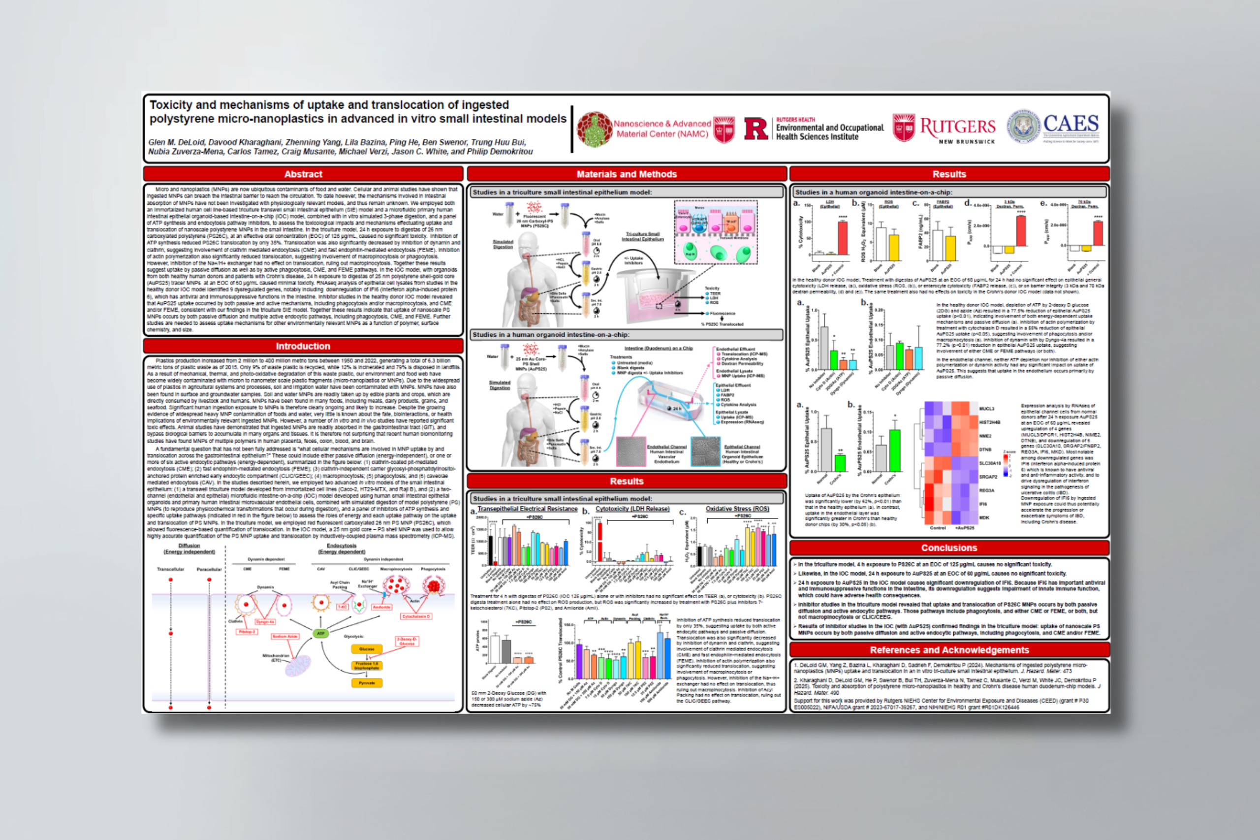
Toxicity and mechanisms of uptake and translocation of ingested polystyrene micro-nanoplastics in advanced in vitro small intestinal models

Originally presented at the Society of Toxicology 2025 Annual Meeting.

Authors

Glen M. DeLoid, Davood Kharaghani, Zhenning Yang, Lila Bazina, Ping He, Ben Swenor, Trung Huu Bui, Nubia Zuverza-Mena, Carlos Tamez, Craig Musante, Michael Verzi, Jason C. White, and Philip Demokritou

Abstract

Micro and nanoplastics (MNPs) are now ubiquitous contaminants of food and water. Cellular and animal studies have shown that ingested MNPs can breach the intestinal barrier to reach the circulation. To date however, the mechanisms involved in intestinal absorption of MNPs have not been investigated with physiologically relevant models, and thus remain unknown. We employed both an immortalized human cell line-based triculture transwell small intestinal epithelium (SIE) model and a microfluidic primary human intestinal epithelial organoid-based intestine-on-a-chip (IOC) model, combined with in vitro simulated 3-phase digestion, and a panel of ATP synthesis and endocytosis pathway inhibitors, to assess the toxicological impacts and mechanisms effectuating uptake and translocation of nanoscale polystyrene MNPs in the small intestine. In the triculture model, 24 h exposure to digestas of 26 nm carboxylated polystyrene (PS26C), at an effective oral concentration (EOC) of 125 μg/mL, caused no significant toxicity. Inhibition of ATP synthesis reduced PS26C translocation by only 35%. Translocation was also significantly decreased by inhibition of dynamin and clathrin, suggesting involvement of clathrin mediated endocytosis (CME) and fast endophilin-mediated endocytosis (FEME). Inhibition of actin polymerization also significantly reduced translocation, suggesting involvement of macropinocytosis or phagocytosis. However, inhibition of the Na+/H+ exchanger had no effect on translocation, ruling out macropinocytosis. Together these results suggest uptake by passive diffusion as well as by active phagocytosis, CME, and FEME pathways. In the IOC model, with organoids from both healthy human donors and patients with Crohn’s disease, 24 h exposure to digestas of 25 nm polystyrene shell-gold core (AuPS25) tracer MNPs at an EOC of 60 μg/mL caused minimal toxicity. RNAseq analysis of epithelial cell lysates from studies in the healthy donor IOC model identified 9 dysregulated genes, notably including downregulation of IFI6 (interferon alpha-induced protein 6), which has antiviral and immunosuppressive functions in the intestine. Inhibitor studies in the healthy donor IOC model revealed that AuPS25 uptake occurred by both passive and active mechanisms, including phagocytosis and/or macropinocytosis, and CME and/or FEME, consistent with our findings in the triculture SIE model. Together these results indicate that uptake of nanoscale PS MNPs occurs by both passive diffusion and multiple active endocytic pathways, including phagocytosis, CME, and FEME. Further studies are needed to assess uptake mechanisms for other environmentally relevant MNPs as a function of polymer, surface chemistry, and size.

Related publication: Toxicity and absorption of polystyrene micro-nanoplastics in healthy and Crohn’s disease human duodenum-chip models

Human Organ-Chips Reveal New Inflammatory Bowel Disease Drivers

Synopsis

In this webinar, Dr. Alican Ozkan from the Wyss Institute discusses how he and his colleagues leveraged human Organ-on-a-Chip technology to model the hallmark features of inflammatory bowel disease (IBD). Using patient-derived colon epithelial cells and matched fibroblasts cultured under flow, the IBD Chips replicated compromised barrier function, decreased mucus accumulation, and heightened risk of inflammation, fibrosis, and cancer—key challenges in IBD patients.

Key highlights from this webinar include how:

  • IBD fibroblasts act as primary drivers of multiple disease hallmarks, including inflammation and fibrosis.
  • Peristalsis-like mechanical forces heighten inflammation and fibrosis within the IBD Chips, reflecting critical in vivo dynamics.
  • Female IBD Chips respond more severely to pregnancy-associated hormones, mirroring clinical observations of aggravated IBD symptoms in pregnant women.
  • Carcinogen exposure increases inflammation, gene mutations, and chromosome duplication exclusively in IBD Chips, underscoring the amplified cancer risk in IBD patients.
  • The human Organ-Chip system reveals the pivotal role of intestinal stroma and mechanical deformations in driving sex-specific disease progression and provides a robust platform for therapeutic testing.

This recording was originally presented on February 24, 2025, and is based on data from this publication.

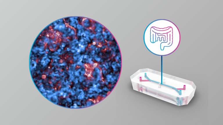
Toxicity and absorption of polystyrene micro-nanoplastics in healthy and Crohn’s disease human duodenum-chip models
Organ Model: Intestine (Duodenum)

Application: ADME, Toxicology

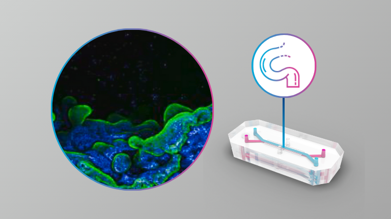
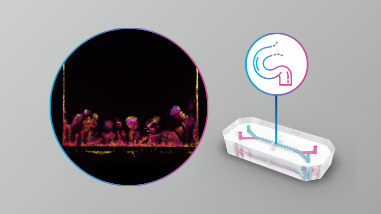
This study used a human Intestine-Chip model and found that 25 nm polystyrene micro/nanoplastics (MNP) caused minimal toxicity in both healthy and Crohn’s-derived small intestinal tissue over 24 hours—no major damage to barrier function or big shifts in inflammatory signals. However, researchers did detect changes in a handful of genes (including IFI6) linked to immune defense. They also confirmed that MNP uptake involves both passive diffusion and active endocytosis pathways—suggesting that even “low-toxicity” levels of plastics can still get absorbed and potentially affect gut biology.

Products Used In This Publication

Inflammatory Bowel Disease Drivers Revealed in Human Organ Chips

Organ Model: Intestine (Colon)

Application: Immunology & Inflammation

How Organ-Chips Were Used: Patient-specific epithelial cells and stromal fibroblasts were isolated and cultured to establish healthy and IBD-specific Colon Intestine-Chips.

Key highlights:

  • Colon Intestine-Chips replicated key hallmarks of IBD, including inflammation, compromised barrier function, reduced mucus accumulation, fibrosis, and increased cancer risk.
  • Stromal fibroblasts from IBD patients were identified as primary drivers of intestinal barrier disruption and inflammation.
  • Peristalsis-like mechanical deformations have a direct effect on mucus production, and they exacerbated inflammation and fibrosis in IBD Chips but not in healthy chips.
  • Mechanical deformations also enhanced immune cell migration in IBD Chips.

Products Used In This Publication

Emulate BioKits: Innovative Case Studies in Disease Modeling and Immunotherapy Safety

In this webinar, Emeli Chatterjee, Postdoctoral Researcher at Massachusetts General Hospital, and Ben Swenor, Senior Science Liaison at Emulate, present case studies from three peer-reviewed papers highlighting how Organ-Chips have been used to study cardiorenal syndrome, environmental enteric dysfunction (EED), and immunotherapy safety. View now to learn how Organ-Chips can be used to investigate complex disease mechanisms and evaluate immunotherapy safety.

Case study topics include: 

  • Cardiorenal Syndrome: Investigating the role of extracellular vesicles from patients with cardiorenal syndrome on renal injury using the Kidney-Chip, offering insights into disease mechanisms and potential therapeutic targets. 
  • Immuno-oncology Safety: Using the Colon Intestine-Chip and Duodenum Intestine-Chip to evaluate the on-target, off-tumor safety of T-cell bispecific antibodies. 
  • Environmental enteric dysfunction (EED): Replicating EED disease mechanisms with the Duodenum Intestine-Chip, comparing the effects of malnutrition with healthy vs. patient-derived tissue to uncover novel therapeutic targets. 

Read the author Q&A here.

Organ chips with integrated multifunctional sensors enable continuous metabolic monitoring at controlled oxygen levels

Organ Model: Small Intestine & Liver

Application: Organ-on-a-Chip Technology

Abstract: Despite remarkable advances in Organ-on-a-chip (Organ Chip) microfluidic culture technology, recreating tissue-relevant physiological conditions, such as the region-specific oxygen concentrations, remains a formidable technical challenge, and analysis of tissue functions is commonly carried out using one analytical technique at a time. Here, we describe two-channel Organ Chip microfluidic devices fabricated from polydimethylsiloxane and gas impermeable polycarbonate materials that are integrated with multiple sensors, mounted on a printed circuit board and operated using a commercially available Organ Chip culture instrument. The novelty of this system is that it enables the recreation of physiologically relevant tissue-tissue interfaces and oxygen tension as well as non-invasive continuous measurement of transepithelial electrical resistance, oxygen concentration and pH, combined with simultaneous analysis of cellular metabolic activity (ATP/ADP ratio), cell morphology, and tissue phenotype. We demonstrate the reliable and reproducible functionality of this system in living human Gut and Liver Chip cultures. Changes in tissue barrier function and oxygen tension along with their functional and metabolic responses to chemical stimuli (e.g., calcium chelation, oligomycin) were continuously and noninvasively monitored on-chip for up to 23 days. A physiologically relevant microaerobic microenvironment that supports co-culture of human intestinal cells with living Lactococcus lactis bacteria also was demonstrated in the Gut Chip. The integration of multi-functional sensors into Organ Chips provides a robust and scalable platform for the simultaneous, continuous, and non-invasive monitoring of multiple physiological functions that can significantly enhance the comprehensive and reliable evaluation of engineered tissues in Organ Chip models in basic research, preclinical modeling, and drug development.

Intestine-on-chip enhances nutrient and drug metabolism and maturation of iPSC-derived intestinal epithelial cells relative to organoids and Transwells

Organ Model: Intestine

Application: ADME

Abstract: The human intestinal epithelial barrier is shaped by various biological and biomechanical influences such as growth factor gradients and the flow of intestinal contents. Exposure to these cues in vitro impacts the cell type composition and function of adult stem cell (ASC)-derived intestinal epithelial cells, but their effect on human induced pluripotent stem cell (hiPSC)-derived cells is largely unexplored. Here, we characterize and compare the cellular composition and gene expression profiles of hiPSC-derived intestinal epithelial cells exposed to various medium compositions and cultured as organoids, in Transwell and in microfluidic intestine-on-chip systems. We demonstrate that inhibition and activation of the WNT, BMP, NOTCH and MAPK pathways regulates the presence of dividing, absorptive and secretory epithelial lineages within these systems, as has been described for ASC-based systems. Upon differentiation, intestinal epithelial organoids and monolayers in Transwell systems expressed genes involved in important intestinal functions, including digestive enzymes, nutrient transporters and members of the Cytochrome P450 family implicated in drug metabolism. However, the dynamic microenvironment of the intestine-on-chip system induced the strongest upregulation of these genes, with an expression profile that suggests a more mature developmental state. Overall, these results underscore the value of hiPSC-derived intestinal epithelial cells for modeling important functions of the human intestinal epithelial barrier and facilitates the selection of relevant culture conditions for specific applications.

Products Used In This Publication

Head-to-Head Comparison of Caco-2 Transwell and Gut-on-a-Chip Models for Assessing Oral Peptide Formulations

Organ Model: Intestine (Caco2)

Application: ADME

How Organ-Chips Were Used: The authors of this publication assessed the utility of a Caco2 Intestine-Chip to study the effect of permeability enhancers (PEs) on the absorption of peptides (Insulin and Octreotide) in the intestine. The authors observed a much more modest permeability enhancement in the Chip model compared to transwell models, which is in line with observations in ex vivo and in vivo preclinical models. These data indicate that microfluidic chip models are well suited to bridge the gap between conventional in vitro and in vivo models.

Products Used In This Publication

An iPSC-derived small intestine-on-chip with self-organizing epithelial, mesenchymal, and neural cells

Organ Model: Small Intestine

Application: Model Development

How Organ-Chips Were Used: In this publication, users developed an hiPSC-derived Intestine-Chip, which includes a diverse epithelial composition with myofibroblast and neural subtypes, as a model for the human small intestine. This well-characterized hiPSC-derived Intestine-Chip system can facilitate personalized studies on physiological processes and therapy development in the human small intestine.

Products Used In This Publication

Clostridioides difficile binary toxin CDT induces biofilm-like persisting microcolonies

Organ Model: Intestine (Caco2)

Application: Infectious Disease

Abstract: Clinical symptoms of Clostridioides difficile infection (CDI) range from diarrhea to pseudomembranous colitis. A major challenge in managing CDI is the high rate of relapse. Several studies correlate production of CDT binary toxin by clinical strains of Clostridioides difficile with higher relapse rates. Although the mechanism of action of CDT on host cells is known, its exact contribution to CDI is still unclear. To understand the physiological role of CDT during CDI, we established two hypoxic relevant intestinal models, Transwell and Microfluidic Intestine-on-Chip systems. Both were challenged with the epidemic strain UK1 CDT+ and its isogenic CDT- mutant. We report that CDT binary toxin induces mucin-associated microcolonies that increase C. difficile colonization and display biofilm-like properties by enhancing C. difficile resistance to vancomycin but not to fidaxomicin, a biofilm disrupting antibiotic. Importantly, biofilm-like CDT-dependent microcolonies were also observed in the caecum and colon of infected mice. Hence, our study shows that CDT toxin induces biofilm-like microcolonies, increasing C. difficile colonization and persistence.