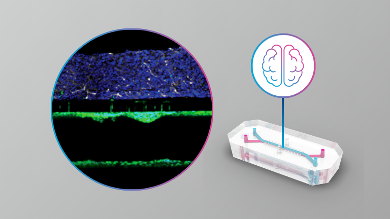
Organ-Chips Enhance the Maturation of Human iPSC-Derived Dopamine Neurons

Organ Model: Brain

Application: Neuroscience

Abstract: While cells in the human body function in an environment where the blood supply constantly delivers nutrients and removes waste, cells in conventional tissue culture well platforms are grown with a static pool of media above them and often lack maturity, limiting their utility to study cell biology in health and disease. In contrast, organ-chip microfluidic systems allow the growth of cells under constant flow, more akin to the in vivo situation. Here, we differentiated human induced pluripotent stem cells into dopamine neurons and assessed cellular properties in conventional multi-well cultures and organ-chips. We show that organ-chip cultures, compared to multi-well cultures, provide an overall greater proportion and homogeneity of dopaminergic neurons as well as increased levels of maturation markers. These organ-chips are an ideal platform to study mature dopamine neurons to better understand their biology in health and ultimately in neurological disorders.

Evaluation of a Human Neurovascular Model to Complement a Parallel Non-human Primate Selection for Blood–Brain Barrier Penetrant AAV Capsids

INTRODUCTION

  • Delivery of genomic medicine to the central nervous system (CNS) is a major hurdle for clinical applications of gene therapy; the blood–brain barrier (BBB) limits the brain distribution of virtually all intravenously administered macromolecules.
  • Several adeno-associated virus (AAV) serotypes, most notably AAV9, distribute to the brain after intravenous (IV) administration but require high doses to achieve limited expression.
  • AAV capsid engineering has produced novel variants that are superior to their parental serotypes and have progressed into the clinic for several indications. However, translation of clinical programs from preclinical models to humans remains a challenge for the entire gene therapy field, including capsid engineering efforts.
    • Two factors for a stringent selection campaign have emerged: library designs that incorporate functional cellular transduction pressure, and selection of appropriate in vitro and/or in vivo models.
  • In this study, we employed SIFTER™ (Selecting In vivo For Transduction and Expression of RNA) to engineer capsids with improved CNS transduction following IV administration in Cynomolgus macaque (non-human primates [NHPs]). This was followed by implementation of an all human cell model of the BBB that recapitulates many key BBB properties to address discordant capsid performance observed in vitro vs in vivo and between species.

Tissue clearing of human iPSC-derived organ-chips enables high resolution imaging and analysis

Organ Model: Spinal cord

Application: Neuroscience

Abstract

Engineered microfluidic organ-chips enable increased cellular diversity and function of human stem cell-derived tissues grown in vitro. These three dimensional (3D) cultures, however, are met with unique challenges in visualization and quantification of cellular proteins. Due to the dense 3D nature of cultured nervous tissue, classical methods of immunocytochemistry are complicated by sub-optimal light and antibody penetrance as well as image acquisition parameters. In addition, complex polydimethylsiloxane scaffolding surrounding the tissue of interest can prohibit high resolution microscopy and spatial analysis. Hyperhydration tissue clearing methods have been developed to mitigate similar challenges of in vivo tissue imaging. Here, we describe an adaptation of this approach to efficiently clear human pluripotent stem cell-derived neural tissues grown on organ-chips. We also describe critical imaging considerations when designing signal intensity-based approaches to complex 3D architectures inherent in organ-chips. To determine morphological and anatomical features of cells grown in organ-chips, we have developed a reliable protocol for chip sectioning and high-resolution microscopic acquisition and analysis.

Products Used In This Publication

A Microengineered Brain-Chip to Model Neuroinflammation in Humans

iScience (2022)

Abstract

Species differences in brain and blood-brain barrier (BBB) biology hamper translation from findings in animal models to humans. This, in turn, impedes the development of medicines and brain diseases. Researchers from Emulate developed a human Brain-Chip that contains endothelial-like cells, pericytes, glia, and cortical neurons while maintaining BBB permeability at in vivo-relevant levels. With these features, the model is able to closely recapitulate neuroinflammation, allowing scientists to gain a mechanistic understanding of cell-cell interactions and BBB function during neuroinflammation with greater accuracy than they could with conventional models of the human brain.

Modeling alpha-synuclein pathology in a human Brain-Chip to assess blood-brain barrier disruption

Nature Communications (2021)

Abstract

Parkinson’s disease and related synucleinopathies are characterized by the abnormal accumulation of alpha-synuclein aggregates, loss of dopaminergic neurons, and gliosis of the substantia nigra. Although clinical evidence and in vitro studies indicate disruption of the Blood-Brain Barrier in Parkinson’s disease, the mechanisms mediating the endothelial dysfunction is not well understood. Here we leveraged the Organs-on-Chips technology to develop a human Brain-Chip representative of the substantia nigra area of the brain containing dopaminergic neurons, astrocytes, microglia, pericytes, and microvascular brain endothelial cells, cultured under fluid flow. Our αSyn fibril-induced model was capable of reproducing several key aspects of Parkinson’s disease, including accumulation of phosphorylated αSyn (pSer129-αSyn), mitochondrial impairment, neuroinflammation, and compromised barrier function. This model may enable research into the dynamics of cell-cell interactions in human synucleinopathies and serve as a testing platform for target identification and validation of novel therapeutics.

Development of a Human Brain-Chip Model to Study Neuroinflammatory Diseases

Abstract

Species differences in brain function and blood-brain barrier often preclude accurate extrapolation from animal models to human patients. There is an unmet need for human relevant systems that can recreate key aspects of brain physiology and pathophysiology of common diseases. We are developing a human Brain-Chip to model neuroinflammation, a hallmark of many neurodegenerative diseases, to enable studies on mechanistic aspects of neural pathology and disease progression. We provide evidence that this complex human Brain-Chip model can support co-culture and establishment of extensive interconnection between human iPSC-derived neurons and primary glia cells (astrocytes and microglia). Human iPSC-derived brain endothelial cells successfully maintained at the vascular channel of the Brain-Chip in the presence of fluidic shear stress, while exhibiting hallmark features of the human blood-brain barrier, such as development of specific tight junctions and minimal barrier permeability. Exposure to inflammatory triggers (e.g. TNF-α) or toxic protein oligomeric species (e.g. alpha-synuclein), resulted in neuronal death, glia activation, increased secretion of the corresponding proinflammatory cytokines, and a compromised barrier function. In summary, our current findings demonstrate the development of a human Brain-Chip that could support the development of models for the study of neuroinflammation and blood-brain barrier disfunction in neurological disorders.

Generation of a Human iPSC-Based Blood-Brain Barrier Chip

Organ Model: Brain (BBB)

Applications: Neuroscience

Abstract: The blood brain barrier (BBB) is formed by neurovascular units (NVUs) that shield the central nervous system (CNS) from a range of factors found in the blood that can disrupt delicate brain function. As such, the BBB is a major obstacle to the delivery of therapeutics to the CNS. Accumulating evidence suggests that the BBB plays a key role in the onset and progression of neurological diseases. Thus, there is a tremendous need for a BBB model that can predict penetration of CNS-targeted drugs as well as elucidate the BBB’s role in health and disease. We have recently combined organ-on-chip and induced pluripotent stem cell (iPSC) technologies to generate a BBB chip fully personalized to humans. This novel platform displays cellular, molecular, and physiological properties that are suitable for the prediction of drug and molecule transport across the human BBB. Furthermore, using patient-specific BBB chips, we have generated models of neurological disease and demonstrated the potential for personalized predictive medicine applications. Provided here is a detailed protocol demonstrating how to generate iPSC-derived BBB chips, beginning with differentiation of iPSC-derived brain microvascular endothelial cells (iBMECs) and resulting in mixed neural cultures containing neural progenitors, differentiated neurons, and astrocytes. Also described is a procedure for seeding cells into the organ chip and culturing of the BBB chips under controlled laminar flow. Lastly, detailed descriptions of BBB chip analyses are provided, including paracellular permeability assays for assessing drug and molecule permeability as well as immunocytochemical methods for determining the composition of cell types within the chip.

Human iPSC-Derived Blood-Brain Barrier Chips Enable Disease Modeling and Personalized Medicine Applications

Organ Model: Brain (BBB)

Application: Neuroscience

Abstract: The blood-brain barrier (BBB) tightly regulates the entry of solutes from blood into the brain and is disrupted in several neurological diseases. Using Organ-Chip technology, we created an entirely human BBB-Chip with induced pluripotent stem cell (iPSC)-derived brain microvascular endothelial-like cells (iBMECs), astrocytes, and neurons. The iBMECs formed a tight monolayer that expressed markers specific to brain vasculature. The BBB-Chip exhibited physiologically relevant transendothelial electrical resistance and accurately predicted blood-to-brain permeability of pharmacologics. Upon perfusing the vascular lumen with whole blood, the microengineered capillary wall protected neural cells from plasma-induced toxicity. Patient-derived iPSCs from individuals with neurological diseases predicted disease-specific lack of transporters and disruption of barrier integrity. By combining Organ-Chip technology and human iPSC-derived tissue, we have created a neurovascular unit that recapitulates complex BBB functions, provides a platform for modeling inheritable neurological disorders, and advances drug screening, as well as personalized medicine.

Human iPSC-Derived Endothelial Cells and Microengineered Organ-Chip Enhance Neuronal Development

Published in: Stem Cell Reports

Abstract

Human stem cell-derived models of development and neurodegenerative diseases are challenged by cellular immaturity in vitro. Microengineered organ-on-chip (or Organ-Chip) systems are designed to emulate microvolume cytoarchitecture and enable co-culture of distinct cell types. Brain microvascular endothelial cells (BMECs) share common signaling pathways with neurons early in development, but their contribution to human neuronal maturation is largely unknown. To study this interaction and influence of microculture, we derived both spinal motor neurons and BMECs from human induced pluripotent stem cells and observed increased calcium transient function and Chip-specific gene expression in Organ-Chips compared with 96-well plates. Seeding BMECs in the Organ-Chip led to vascular-neural interaction and specific gene activation that further enhanced neuronal function and in vivo-like signatures. The results show that the vascular system has specific maturation effects on spinal cord neural tissue, and the use of Organ-Chips can move stem cell models closer to an in vivo condition.