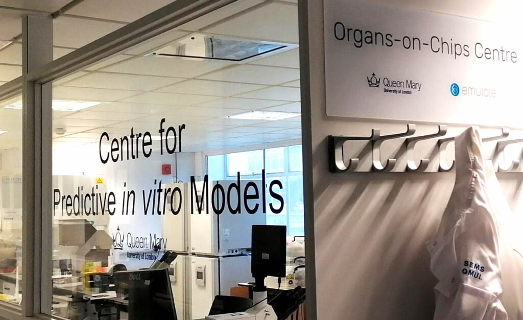
For academic researchers, keeping up with innovative approaches to solving problems can be a never-ending exercise as new technologies displace older methods of experimentation. At Queen Mary University London (QMUL), scientists across many disciplines are using in vitro models to explore the fundamental biology of health and disease and to develop and test novel therapeutic approaches, such as pharmaceuticals, regenerative medicine techniques, medical devices, and biomaterials. QMUL’s Centre for Predictive in vitro Models (CPM) opened in 2019, providing multidisciplinary research focusing on the development and use of predictive in vitro models. This covers a wide range of model systems including 2D and 3D cell culture models, organoids, microphysiological systems, organ-on-a-chip technology, and other types of in vitro models.The vision of the CPM is to facilitate the implementation of innovative in vitro models in orderto deliver the highest quality discovery science and transformative healthcare solutions. Within the CPM, the University has established the Queen Mary+Emulate Organs-on-Chips Centre. This provides access to Emulate’s platform technology and associated 2D (2 Dimensional) and 3D cell culture models for staff at QMUL and across the wider UK Organ-on-a-Chip Technology Network.
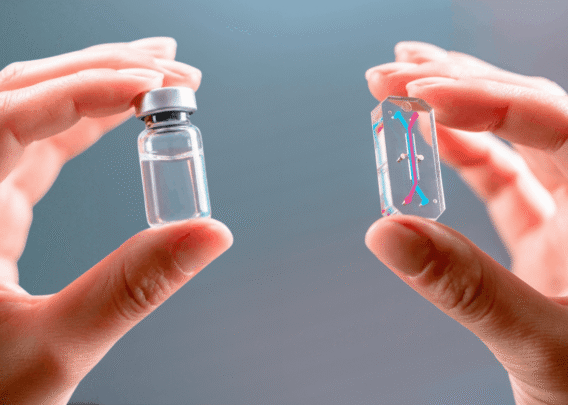
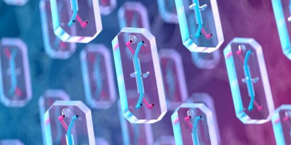
Organs-on-Chips can predict human response with greater precision and control than cell culture or animal-based testing. As such, they are used to test therapeutics and better understand how the body works. For scientists who want to move from static culture models to dynamic organ-chip technology, the QM+Emulate Organs-on-Chips Centre provides access to and support for Emulate Organ-chips. With a project in mind, researchers can perform experiments using Emulate’s established Organ-Chip models, including Brain, Liver, Proximal Tubule Kidney, and Intestine, or they can develop models of their own design using Emulate’s Basic Research Kit. Emulate’s technology can be configured to recreate a wide range of different organ tissues and disease models. They recreate the body’s dynamic cellular microenvironment, including tissue interfaces, blood flow, immune cell interactions, and mechanical forces. Staff at the Centre provide expert-level training on the platform and assistance with experimental design for proof-of-concept studies.
Modeling Bone Metastasis with Emulate Organ-Chips
Much of the work being done at the QM+Emulate Centre using Organ-Chips is breaking new ground for in vitro modeling and providing unprecedented biological insight. In many experimental situations, two-dimensional cell culture methods are unable to replicate the appropriate cellular microenvironment, and animal models are unsuitable due to species differences. One example of current Organ-Chip research at Queen Mary involves the developed breast cancer bone metastasis model. This provides a model of mechanically active bone tissue that enabled analysis of cancer cell migration, proliferation, and invasion.
This research, funded by Cancer Research UK, the Engineering & Physical Sciences Research Council (EPSRC), and the EU, is being led by Prof. Martin Knight’s group based in the Bioengineering department within the School of Engineering and Materials Science. The group recently published a paper which sought to understand if the osteocyte environment, which includes mechanical stimulation via fluid shear stress, can regulate the behavior of breast and prostate cancer cells. The research conducted by Dr. Stefaan Verbruggen discovered that osteocytes signaled for decreased proliferation of cancer cells, but mechanical loading reversed this phenomenon in breast cancer cells. With the help of the microfluidic Organ-Chip model, the authors demonstrated the importance of replicating the mechanical tumor environment, which led to increased invasion potential of cancer cells. Understanding this mechanism will help the research team develop new treatments for breast and prostate cancer bone metastasis, which currently have very poor survival rates.
Excitement about Organ-Chips is Building in the UK
Research successes, like modeling bone metastasis, are generating great interest in in vitro predictive model systems in the UK. “We are establishing proof-of-concept programs to enable researchers to get started with Organ-Chip models and are currently supporting more than 20 groups building new models including tendon, bone, cartilage, placenta, and synovium in addition to developing new protocols for Emulate’s existing models,” said Dr. Clare Thompson, Emulate Centre Scientist. “Many are really drawn to the imaging capabilities of the chip models. In addition to viewing fixed chips with confocal microscopy, you can perform live imaging studies to look at how your cells are behaving in real time- this is always quite exciting for our researchers.”
Since its opening, the QM+Emulate Organs-on-Chips Centre has supported a variety of grant applications for researchers at Queen Mary University and other institutions. Research topics range from testing new cancer therapeutics to modeling immune cell interactions to asking basic questions in biology and physiology that examine how mechanical forces impact organs and cells. The Centre also provides opportunities for collaboration with Emulate as well as support for commercialization and translational impact.
For more information on getting started with Organ-Chips at QMUL, visit https://www.cpm.qmul.ac.uk/