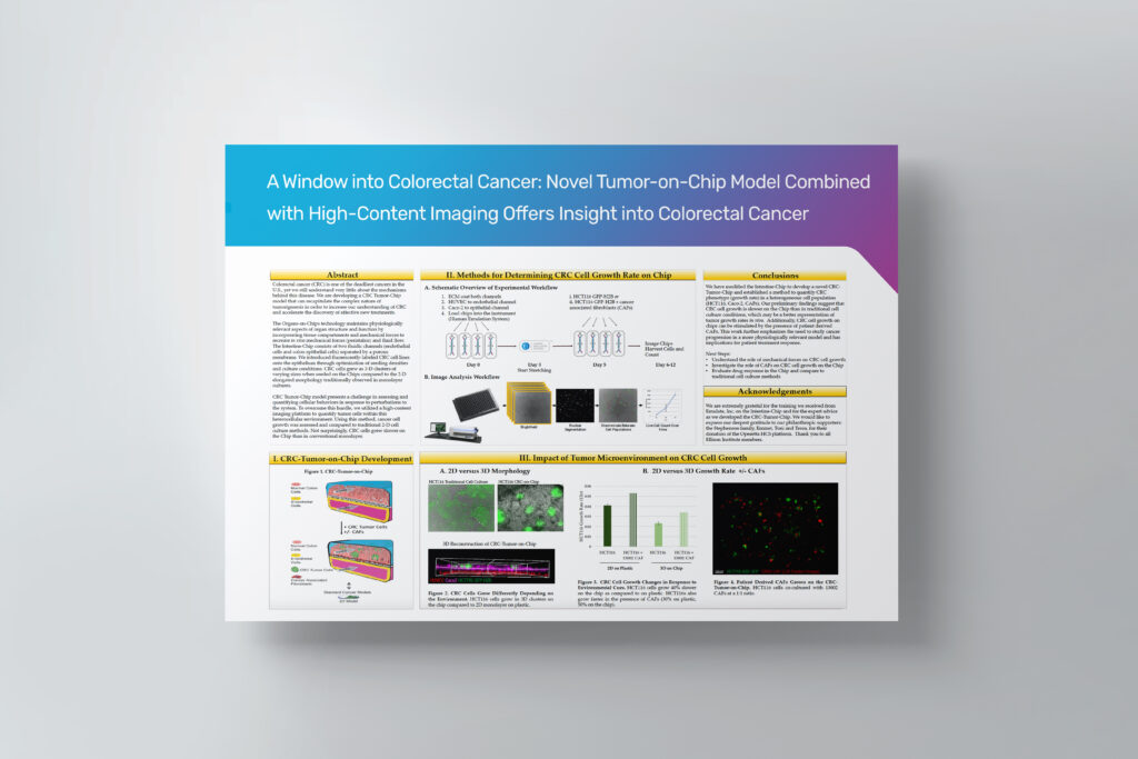
Abstract
Colorectal cancer (CRC) is one of the deadliest cancers in the U.S., yet we still understand very little about the mechanisms behind this disease. We are developing a CRC Tumor-Chip model that can recapitulate the complex nature of tumorigenesis in order to increase our understanding of CRC and accelerate the discovery of effective new treatments.
The Organs-on-Chips technology maintains physiologically relevant aspects of organ structure and function by incorporating tissue compartments and mechanical forces to recreate in vivo mechanical forces (peristalsis) and fluid flow. The Intestine-Chip consists of two fluidic channels (endothelial cells and colon epithelial cells) separated by a porous membrane. We introduced fluorescently-labeled CRC cell lines onto the epithelium through optimization of seeding densities and culture conditions. CRC cells grew as 3-D clusters of varying sizes when seeded on the Chips compared to the 2-D elongated morphology traditionally observed in monolayer cultures.
CRC Tumor-Chip model presents a challenge in assessing and quantifying cellular behaviors in response to perturbations to the system. To overcome this hurdle, we utilized a high-content imaging platform to quantify tumor cells within this heterocellular environment. Using this method, cancer cell growth was assessed and compared to traditional 2-D cell culture methods. Not surprisingly, CRC cells grew slower on the Chip than in conventional monolayer.