Methods
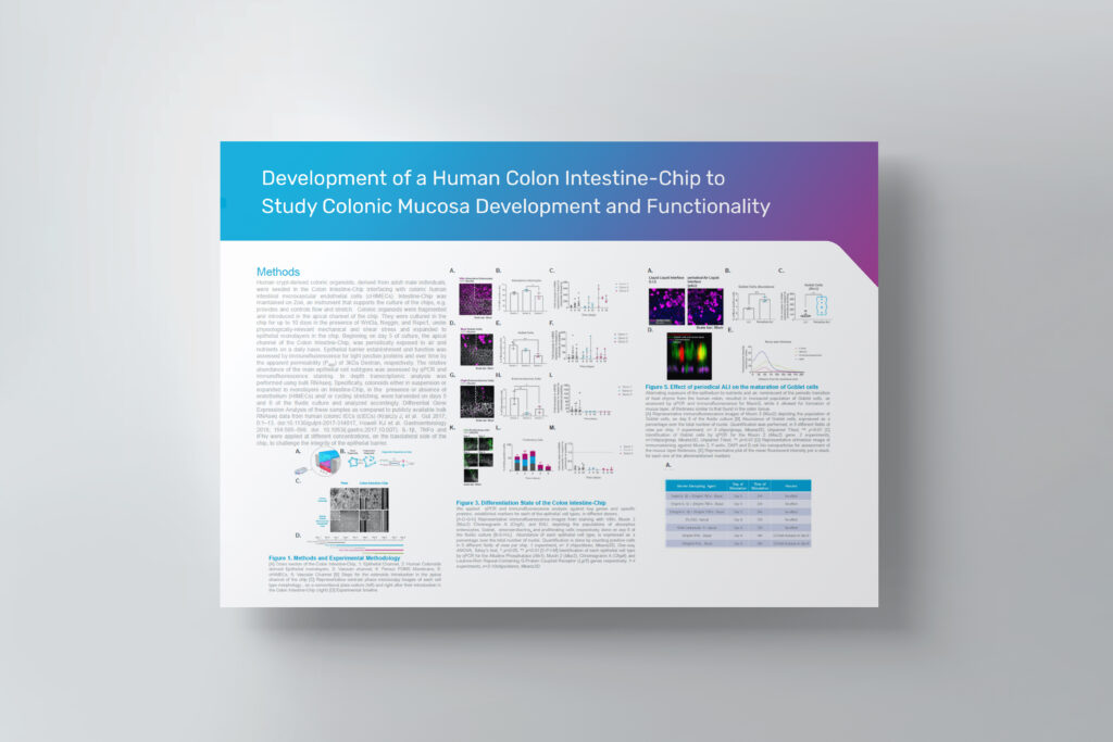
Human crypt-derived colonic organoids, derived from adult male individuals, were seeded in the Colon Intestine-Chip interfacing with colonic human intestinal microvascular endothelial cells (cHIMECs). Intestine-Chip was maintained on Zoë Culture Module, an instrument that supports the culture of the chips, e.g. provides and controls flow and stretch. Colonic organoids were fragmented and introduced in the apical channel of the chip. They were cultured in the chip for up to 10 days in the presence of Wnt3a, Noggin, and Rspo1, under physiologically-relevant mechanical and shear stress and expanded to epithelial monolayers in the chip. Beginning on day 5 of culture, the apical channel of the Colon Intestine-Chip, was periodically exposed to air and nutrients on a daily basis. Epithelial barrier establishment and function was assessed by immunofluorescence for tight junction proteins and over time by the apparent permeability (Papp) of 3kDa Dextran, respectively. The relative abundance of the main epithelial cell subtypes was assessed by qPCR and immunofluorescence staining. In depth transcriptomic analysis was performed using bulk RNAseq. Specifically, colonoids either in suspension or expanded to monolayers on Intestine-Chip, in the presence or absence of endothelium (HIMECs) and/ or cycling stretching, were harvested on days 5 and 8 of the fluidic culture and analyzed accordingly. Differential Gene Expression Analysis of these samples as compared to publicly available bulk RNAseq data from human colonic IECs (cIECs) (Kraiczy J, et al. Gut 2017; 0:1–13. doi:10.1136/gutjnl-2017-314817, Howell KJ et al. Gastroenterology 2018; 154:585–598. doi: 10.1053/j.gastro.2017.10.007). IL-1β, TNFα and IFNγ were applied at different concentrations, on the basolateral side of the chip, to challenge the integrity of the epithelial barrier.

